Popular Microbiology Labs

Find the labs teachers run most often, what students do in each, the results to expect, and the kits and supplies that support them. Procedures for several of these labs are in our free guide, Techniques for Studying Bacteria and Fungi.

Basic Techniques

Start here. These labs build the handling and observation skills that every other lab on this page depends on.

Aseptic technique

Aseptic technique is the foundation of all work with microorganisms and biological materials. It keeps cultures and experiments free from contamination, which protects the accuracy of your results. It also protects the experimenter and the environment by keeping harmful organisms from spreading.

Introduction to Sterile Technique

Students practice transferring a bacterial culture using sterile technique. This is an introductory activity; make sure students already know standard lab procedures, including gas burner use and PPE.

sterile technique

Isolating Bacteria from a Mixture

Students are introduced to a basic technique used in the study of bacteria: isolating a single species of bacterium from a mixture. In this activity, students practice the technique by using a mixture of bacteria of known composition.

bacillus subtilis (rods)

Bacterial Morphology

Students view cell shapes on prepared slides, or prepare their own slides and simple stains with living cultures. Either way, they need a microscope with oil immersion. To combine environmental sampling with colony identification, use the Bacterial Investigative BioKit.

Bacterial Staining

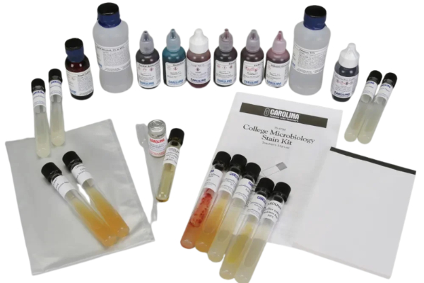
Simple stains add contrast; differential stains sort bacteria into groups based on cell wall differences. The Gram stain divides most bacteria into Gram-positive (blue) and Gram-negative (pink). The College Microbiology Stain Kit covers Gram, acid-fast, endospore, and capsule stains.

Bacterial genetics

Gene transfer between bacteria underpins many biotechnology tools, and it is one way antibiotic resistance spreads.

Bacterial Conjugation

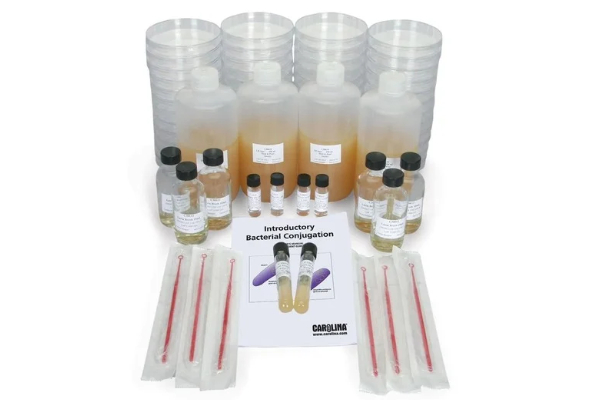
Students confirm strains with different antibiotic resistance on selective media, combine them on a mating plate, then test the results to show that DNA moved between two genetically different cells.

Biochemical identification

Biochemical tests tell apart organisms that look alike under the microscope. Each test below lists the results students should expect.

Motility and SIM

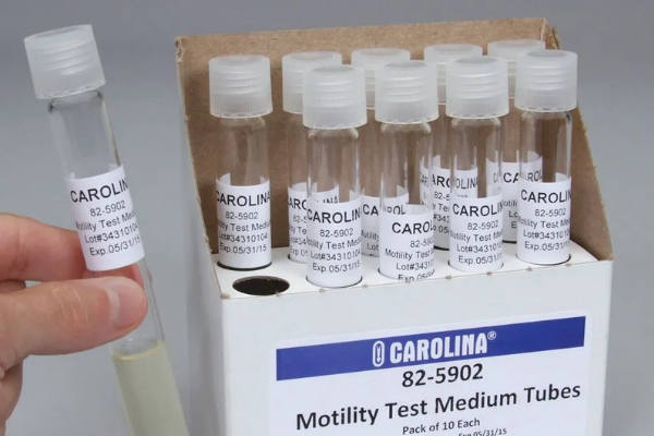
Test motility with the hanging-drop method (depression slide and microscope) or by stabbing motility medium. SIM medium adds sulfide production and indole formation in the same tube.

Biochemical tests of bacteria

OrganismMotilitySulfideIndole
Bacillus cereus+ positive− negative− negative
Escherichia coli+ positive− negative+ positive
Enterobacter aerogenes+ positive− negative− negative
Proteus mirabilis+ positive+ positive− negative
Salmonella typhimurium+ positive+ positive− negative
Serratia marcescens+ positive− negative− negative

The IMViC series

Indole, methyl red, Voges-Proskauer, and citrate are run together to sort common enteric bacteria. Results for all four are combined in one table.
OrganismIndoleMethyl redVoges-ProskauerCitrate
Escherichia coli+ positive+ positive− negative− negative
Enterobacter aerogenes− negative− negative+ positive+ positive
Serratia liquefaciens—+ positive+ positive—
Citrobacter freundii———+ positive
Klebsiella pneumoniae———− negative

Indole test

Kovac’s reagent turns red where bacteria have broken tryptophan down into indole, and stays yellow where they have not.

Methyl red / Voges-Proskauer

MR detects stable acid end products from mixed-acid fermentation; VP detects acetoin from the butylene glycol pathway.

Citrate test

The citrate test determines if organisms can use citric acid as their sole carbon source. 

Carbohydrate fermentation (Durham tubes)

Phenol red broth turns yellow when an organism produces acid from the sugar. A bubble in the inverted Durham tube shows that gas was produced.

Fermentation results by sugar

OrganismDextroseLactoseMaltoseMannitolSucrose
Bacillus cereus+ positive− negative+ positive− negativevariable
Proteus mirabilis+ positive− negative− negative− negative− negative
Salmonella typhimurium+ positive− negative+ positive+ positive− negative
Serratia marcescens+ positive− negative+ positive+ positive+ positive
Staphylococcus epidermidis+ positive+ positive+ positive− negative+ positive

Blood agar hemolysis

Complete breakdown of hemoglobin leaves a clear zone around the colonies; partial breakdown leaves a greenish zone.

Hemolysis on blood agar

OrganismHemolysis
Streptococcus pneumoniaeAlpha (incomplete)
Streptococcus pyogenesBeta (complete)
Staphylococcus epidermidisGamma (none)

Kits that combine several tests

Ready to run several tests at once? These kits bundle the media and organisms.

Enteric Biochemical Detective Kit

Citrate use, hydrogen sulfide production, butanediol fermentation, and mixed-acid fermentation.

Bacterial Biochemical Identification Kit

Starch, lipid, and protein degradation tests to tell apart 3 Bacillus species.   

Selective and differential media

Each medium below both limits which organisms grow and shows a visible difference between the ones that do.

MacConkey agar

Students confirm strains with different antibiotic resistance on selective media, combine them on a mating plate, then test the results to show that DNA moved between two genetically different cells.

Growth on MacConkey agar

OrganismGram stainLactose fermentation
Bacillus subtilisGram +No growth
Escherichia coliGram −+ positive
Pseudomonas fluorescensGram −− negative

Mannitol salt agar

High salt favors salt-tolerant organisms. Mannitol fermenters turn the phenol red indicator yellow.

Growth on mannitol salt agar

OrganismMannitol fermentation
Staphylococcus aureus+ positive
Staphylococcus epidermidis− negative

Eosin methylene blue (EMB) agar

Selective for Gram-negative bacteria. Strong lactose fermenters such as E. coli form purple colonies, often with a green metallic sheen; weak fermenters form pink colonies.

Growth on EMB agar

OrganismGram stainLactose fermentation
Bacillus subtilisGram +No growth
Escherichia coliGram −+ positive (strong)
Enterobacter aerogenesGram −+ positive (weak)
Pseudomonas fluorescensGram −− negative

Medical microbiology

Bring real infectious disease testing into the classroom safely.

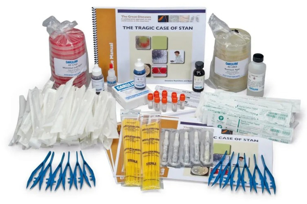
The Tragic Case of Stan

A laboratory case study developed with Tufts University’s Center for Translational Science Education. Students work through a real medical problem facing hospitals around the world.

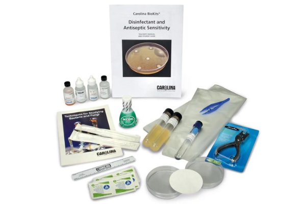
Antiseptic vs. Disinfectant Sensitivity

Students compare how well 5 common disinfectants and antiseptics work against Escherichia coli (Gram-negative) and Bacillus cereus (Gram-positive).

Food microbiology

People have controlled food spoilage in many ways. These kits test how salt, sugar, spices, and chemical preservatives affect microbial growth.

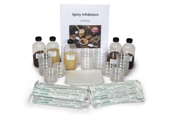
Spicy Inhibitors

Tests how common spices affect microbial growth.

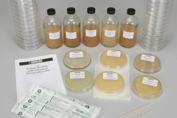
Foiling Spoilage with Chemical Preservatives

Tests how chemical preservatives slow spoilage.

Stocking a new microbiology lab?

The Microbiology Buying Guide covers safety, PPE, media, and equipment in one place.

Newsletter Signup

Get the latest news, free activities, teacher tips, product info, and more delivered to your inbox.

Please login or register to save your favorite articles and activities to your library!