Mitosis is a process ubiquitous to life, and a foundational topic necessary for the understanding of advanced topics like growth and development, reproduction, and many disease processes. However, students are often completely unfamiliar with mitosis. A comprehensive lesson plan for covering the topic is critical for building a foundation for student success in your biology course.
This guide breaks down the important information students need to know about mitosis, provides links to free digital resources for mitosis, and includes suggestions for hands-on-labs that reinforce student learning. Links to helpful worksheets and a quiz are provided.
Prior Knowledge
Every state has standards related to cellular division in its high school biology course of study. As an example, the Next Generation Science Standards* (NGSS) include the following performance expectations:
Students who demonstrate understanding can:
|
|
Use a model to illustrate the role of cellular division (mitosis) and differentiation in producing and maintaining complex organisms. [Assessment Boundary: Assessment does not include specific gene control mechanisms or rote memorization of the steps of mitosis.] |
|
HS-LS3-1. |
Ask questions to clarify relationships about the role of DNA and chromosomes in coding the instructions for characteristic traits passed from parents to offspring. [Assessment Boundary: Assessment does not include the phases of meiosis or the biochemical mechanism of specific steps in the process.] |
|
HS-LS3-2. |
Make and defend a claim based on evidence that inheritable genetic variations may result from (1) new genetic combinations through meiosis, (2) viable errors occurring during replication, and/or (3) mutations caused by environmental factors. [Clarification Statement: Emphasis is on using data to support arguments for the way variation occurs.] [Assessment Boundary: Assessment does not include the phases of meiosis or the biochemical mechanism of specific steps in the process.] |
The Cell Cycle
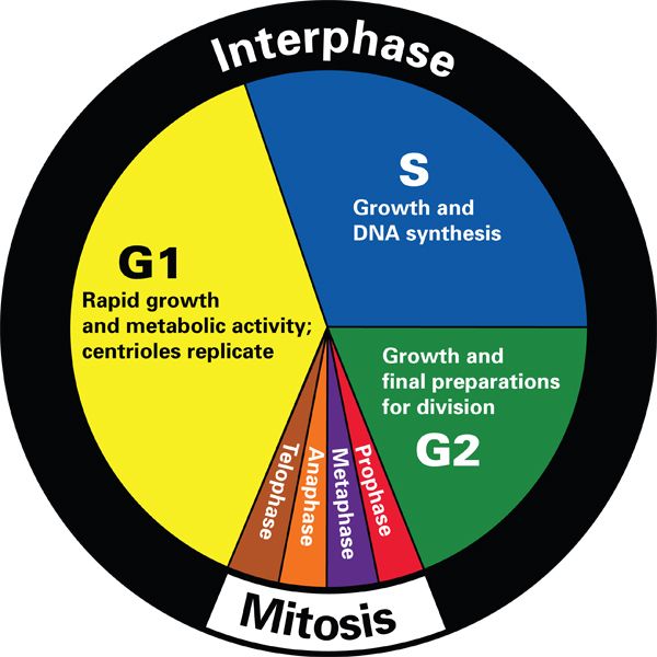
Figure 1 – Representation of the phases of the cell cycle: gap phase 1, synthesis phase, gap phase 2, and mitosis.
The cell cycle can roughly be separated into 2 parts: interphase and mitosis. The mitotic phase is the shortest part of the cell cycle. The rest of the cell cycle, interphase, accounts for about 90% of the cell cycle. Both interphase and mitosis can be broken down into more discrete phases.
A common misconception is that the cell cycle and mitosis are synonymous. Mitosis is just one part of the cell cycle. Most cells spend much of their time in interphase, a period of growth, rest, and DNA synthesis.
View “Phases of the cell cycle” to learn more in-depth information
Phases of the Cell Cycle
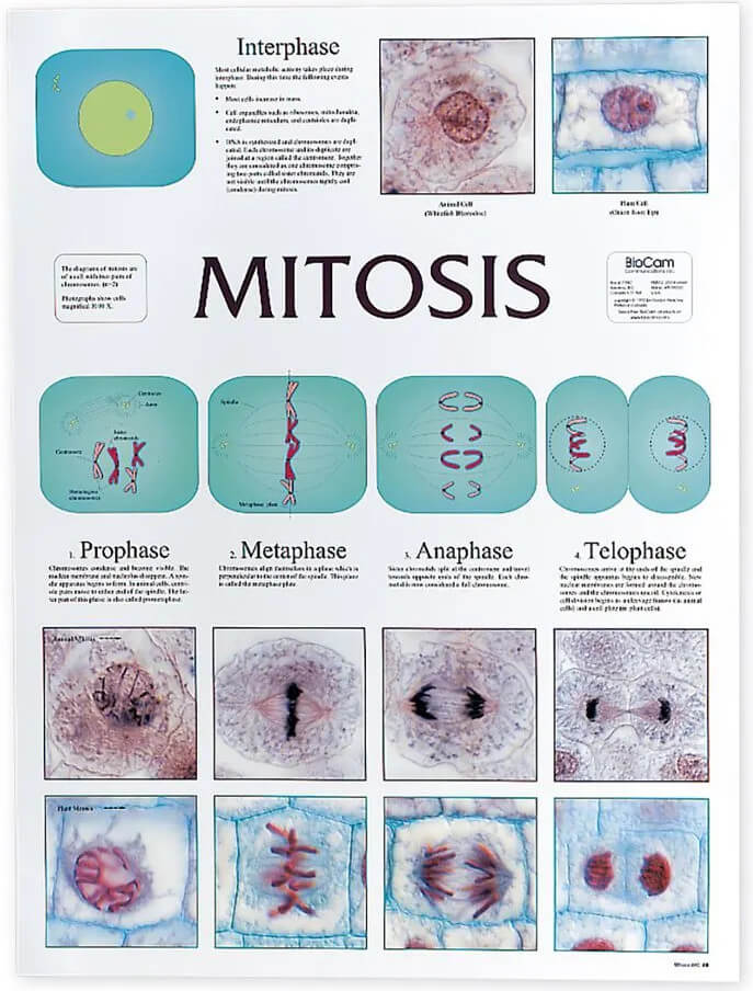
Interphase
Interphase was once referred to as the “resting phase” of the cell cycle; we now know this statement is incorrect. During interphase the cell is busy. The cell grows in size, new organelles are made, and the DNA of the chromosomes are copied in preparation for mitosis and cytokinesis. Interphase is subdivided into 3 phases: G1, S, and G2.
Stages of Interphase:
G1 or gap 1 phase
During the gap 1 (G1) phase, the cell grows larger and the number of organelles increases.
S phase or synthesis phase
During S phase, the cell replicates its DNA. DNA replication only occurs if the cell is programed to proceed beyond G1. At this point the cell possesses twice as much DNA as it normally does and will need to divide. This video from the DNA Learning Center explains the process.
G2 or gap 2 phase
After more growth occurs in the Gap 2 (G2) phase, the cell is ready to divide and enters mitosis.
Mitosis
What is mitosis?
Mitosis is a type of nuclear cell division that occurs in somatic (non-reproductive) eukaryotic cells. At the end of mitosis, the new daughter cells contain the same number of chromosomes as the parent cell. Mitosis enables cellular growth and repair in multicellular organisms.
Cell division differs between plants and animals. For a thorough understanding of the process students should observe mitosis in both plant and animal cells. A chart illustrating the differences in mitosis between plant and animal cells allows students to visualize the difference side by side.
NOTE: Prokaryotic cells reproduce through binary fission where the cell copies its DNA and then splits into 2 new cells.
Mitotic Phases
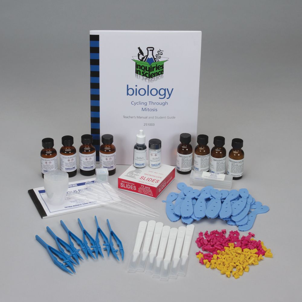
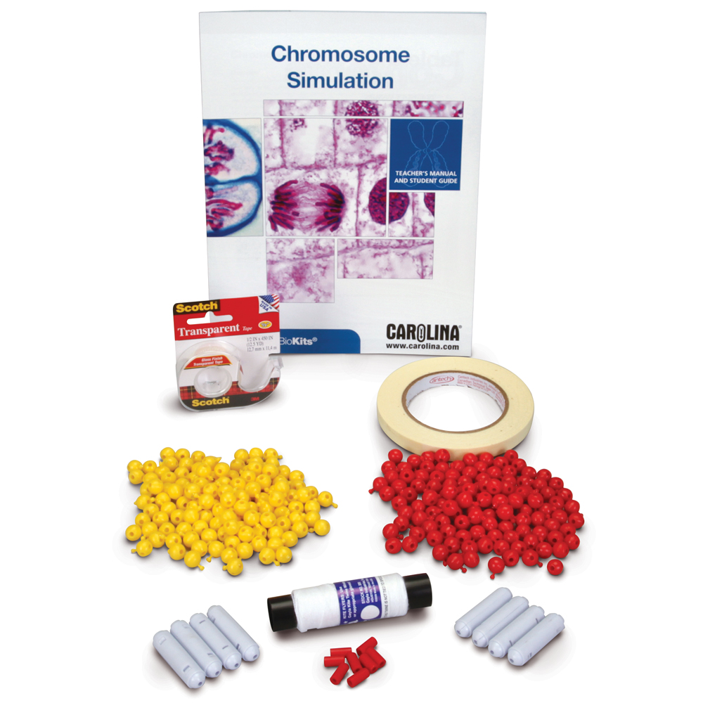
Mitosis takes place in 4 distinct yet continuous phases: prophase, metaphase, anaphase, and telophase. Cytokinesis overlaps with the later stages of mitosis to compete the mitotic phase. The mitotic phase results in the formation of 2 genetically identical daughter cells. Modeling the steps of mitosis is a great way to teach students what takes place in each phase. This activity is covered in Inquires in Science®: Cycling Through Mitosis. For a more in-depth look at chromosomes and their roles in mitosis and meiosis, including more complex events such as crossing over, see Carolina Biokits®: Chromosome Simulation. It allows students to model the process with pop-it beads and magnetic centromeres.
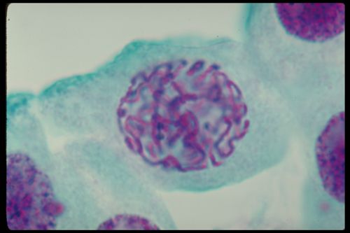
Prophase
In the first phase of mitosis:
- Chromatin coils tightly into visible chromosomes, each with 2 identical sister chromatids joined by the centromere.
- The nuclear envelope begins to break down.
- In animal cells, centrioles separate and move to opposite poles of the cell.
Metaphase
During the second mitotic phase:
- The nuclear envelope is completely gone, allowing for the formation of a spindle fiber.
- Chromosomes line up along the equator, with spindle fibers providing the framework that holds them in place.
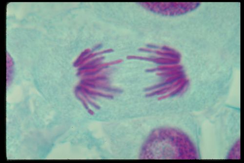
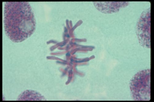
Anaphase
Anaphase is the shortest phase of division.
- Chromosomes are separated into individual chromatids that are pulled to opposite poles.
- Cytokinesis begins.
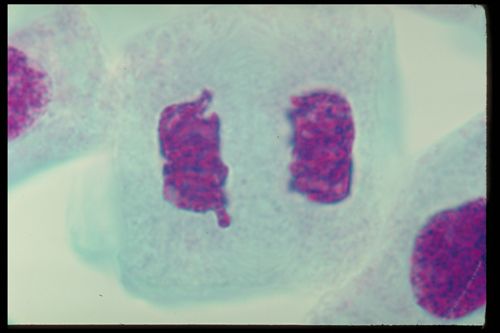
Telophase
During the final mitotic phase:
- The recently separated chromatids uncoil into the chromatin, as seen in interphase.
- Nuclear envelopes reform around the chromatin on each side of the dividing cell.
Cytokinesis
Not an actual stage of mitosis, cytokinesis begins in late anaphase. During this process the cytoplasm begins to separate. In animal cells, the cell membrane simply pinches at the equator of the cell until 2 new cells are formed.
Plant cells, however, have a cell wall, and cannot easily pinch into 2 cells. Instead, tiny vesicles from the Golgi apparatus line up along the equator to form a cell plate. These vesicles fuse forming 2 daughter cells with new cell membranes. Cellulose deposits form between the new membranes, creating new cell walls between the 2 daughter cells. The new cells enter interphase and continue through the cell cycle.
Control of the Cell Cycle
Not all cells are programmed to divide. There are checkpoints at different stages of the cell cycle. Some cells, such as nerve cells, do not continue past the G1 checkpoint of interphase, do not enter synthesis, and therefore do not enter mitosis under normal conditions. Other cells, such as skin cells, are continually undergoing division.
Sometimes genes mutate so that the normal checkpoints are either ignored or overridden. This results in rapid, uncontrolled cell growth. Cancer is an example of a disease caused by loss of cell cycle control. Some cancers are inherited when mutated genes are passed from one generation to the next. Others occur when mutagens, such as UV light, alter a normal cell’s DNA.
Students can learn more about mutation accumulation in cells progressing toward cancer in Collecting Cancer-Causing Changes Kit.
The infographic “Genomics-Driven Oncology” provides extensive information about the role of genomics and mutations in different types of cancers.
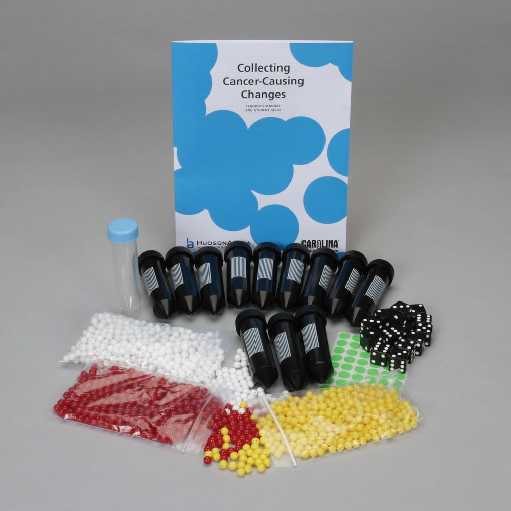
This kit, from the HudsonAlpha Institute of Biotechnology, uses a series of videos that include discussion of cutting-edge research and treatments in oncology along with an activity that demonstrates that a predisposition to the disease along with some chance mutations can lead to cancer.
Mitosis Labs and Classroom Activities
When it comes to learning, nothing beats the hands-on experiences of lab activities. Carolina offers a wide range of free resources, materials, and kits to meet your lab needs.
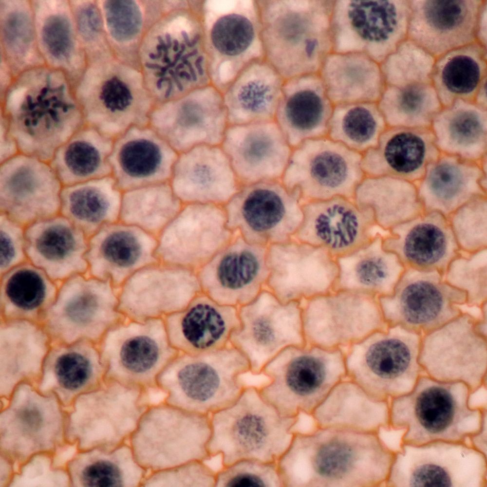
Cell cycle phase investigation
Students can directly observe and calculate the amount of time different cell types spend in interphase and the phases of mitosis by counting the number of cells in each phase. In the kit Inquires in Science®: Cycling Through Mitosis, students design and conduct an investigation to determine the relative lengths of the phases of the cell cycle. The kit includes additional activities relating to mitosis for a comprehensive lesson on the topic.
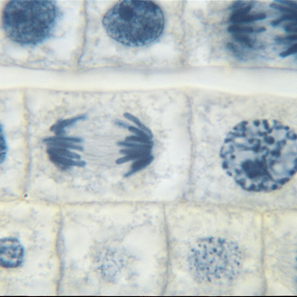
Onion root tip mitosis
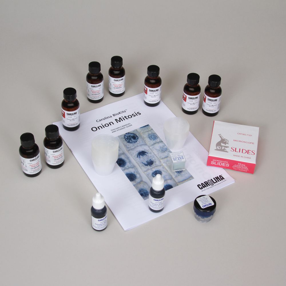
Onion root tips are an excellent model for studying mitosis and the cell cycle. Large cells make for easy viewing by students. The root tip is a fast growth area of the onion plant; cells are rapidly dividing there. That division is not synchronized, so students can observe cells in various phases of the cycle. You can use this guide to prepare your own onion root tip slides or purchase prepared onion root tip slides to perform the activity. Carolina BioKits®: Onion Mitosis offers all the supplies needed to prepare these types of slides.
Related Items
If you have digital microscopes in your classroom or cameras for your microscopes, students can capture the images of the cells in their microscope to use for lab reports, study, or even a presentation to the class.
Modeling mitosis
Activities where students model the process of mitosis are excellent for the kinesthetic learners in your class. Carolina offers these cell model options:
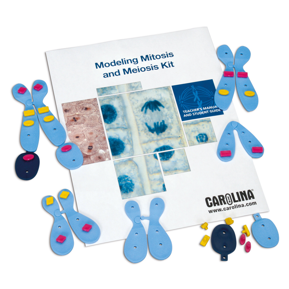
Modeling Mitosis and Meiosis Kit
This kit, covers mitosis, chromosome replication, meiosis, and genetic inheritance using representative autosomes and sex chromosomes.
Chromosome Simulation Kit
This kit, allows students to build their own chromosomes using pop-it beads and magnetic chromosomes to model mitosis and meiosis as well as the more complex topics of crossing over and chromosome aberrations.
Modeling Mitosis and Meiosis Kit covers mitosis, chromosome replication, meiosis, and genetic inheritance using representative autosomes and sex chromosomes.
Chromosome Simulation Kit allows students to build their own chromosomes using pop-it beads and magnetic chromosomes to model mitosis and meiosis as well as the more complex topics of crossing over and chromosome aberrations.
Mitosis Videos and Other Digital Resources
Whether you have a traditional classroom or have adopted a flipped methodology, digital assets are a critical piece for teaching most science concepts. Many of the kits from Carolina now come with digital resources to enhance the learning experience for students.
Our Mitosis Interactive Whiteboard Exercise teaches students about the stages of mitosis in both plant and animal cells while assessing their understanding of the concepts. The DNA Learning Center has a library of exceptional 3-D animations, including videos about DNA packaging into chromosomes.
Quizzes and Assessments
Misconceptions about the cell cycle are common, and therefore frequent checks of student understanding are crucial. By the end of the lessons on this concept, students should be able to answer the following questions:
- Where does mitosis occur?
- Why is mitosis important?
- How does mitosis differ in plant and animal cells?
- What happens during each phase of the cell cycle? Illustrate the steps.
- What is the role of DNA in the cell cycle?
- What changes occur in chromosomes during the cell cycle?
- What happens if there are errors in DNA coding during the cell cycle?
You can use this mitosis worksheet to test students’ knowledge of the phases of mitosis and this interactive whiteboard activity for group assessment.