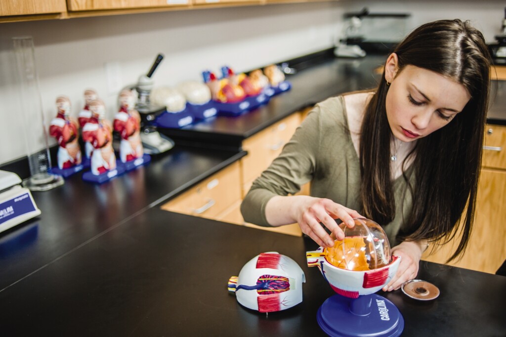
Models are among the most traditional and useful items in a science educator’s toolbox. Whether your subject of study is leaf morphology, invertebrate zoology, general biology, or human anatomy, models are the perfect adjunct to texts and diagrams. They enable students to examine the finest structural details of an organism or its components in 3-dimensonal perspective.
In many cases, models can be disassembled in a manner similar to performing a dissection. As a supplement to dissection, models are an excellent reference tool for identifying small and indistinct structures. With the careful planning and execution of the master sculpture (from which the mold is made), high-quality models display the maximum number of anatomical structures.
Human anatomy models
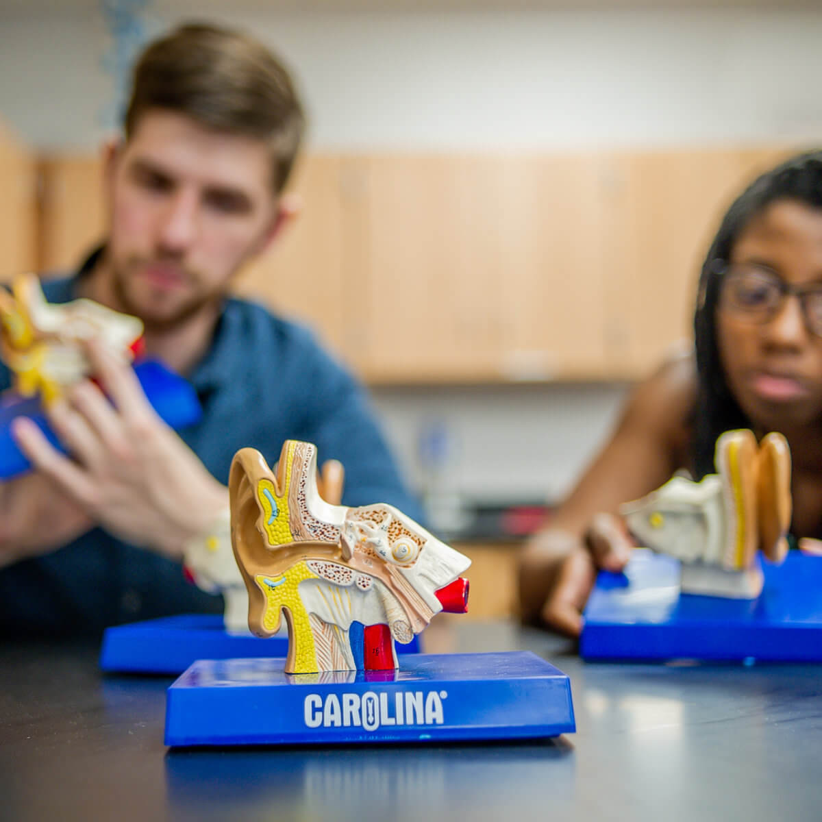
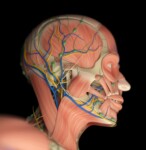
No discipline of science relies more on the use of anatomical models than human anatomy and physiology. Preserved human tissues are not readily available for educational purposes, and when they are, the condition of the specimens is usually not satisfactory to show detailed morphology. Many structures, such as the components of the middle and inner ear, are much too small for convenient study from natural specimens.
A thorough knowledge of the structure of cells and tissues is an absolute prerequisite for an understanding of the physiology, or function, of the various organ systems. An enlarged model is an indispensable teaching aid in such instances.
Which models should I have in my classroom?
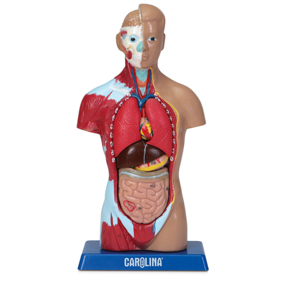
Human torso models
A complete human figure or torso model with head is actually a collection of models of individual organs, many of which can be separated and even opened for internal inspection. A human torso model of high quality is a valuable acquisition for any anatomy classroom. Generally, those with the largest number of removable components have the greatest teaching value.
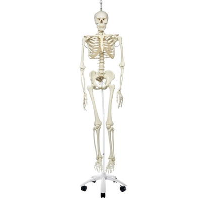
Skeleton models
A staple in many science classrooms, skeletons allow students to examine bones without other organs obstructing their view. Disarticulated skeletons are perfect for learning about the shapes and structure of bone; they are also great for forensic studies. Muscular skeletons have origin and insertion points indicated on the bones to help students understand how the skeletal and muscular systems work together to gain an appreciation for movement. Flexible and ligamentous skeletons offer students an opportunity to visualize the movement of the skeletal system and the articulation of joints.
Organ and organ system models
For a detailed look at specific organs and structures, consider organ and organ system models. These provide a more focused view of the major anatomical structures within systems like the respiratory and urinary systems. Organ models are often made of many pieces, making it easy to explore the internal structures as well.
Disease and pathology models
Providing examples of disease and pathology for students is paramount to understanding the structure and function of human tissue. Tissue pathology models give students an opportunity to observe enlarged examples of diseased tissue, with aids in their ability to clearly identify similar features under a microscope. Organ pathology and disease models, including this hypertension model, show how disease affects the entire system or body.
Care of models
Models represent a formidable investment, and with reasonable care, they will last for many years. Encourage your students to handle the models, but to do so with care and respect, particularly when removing or replacing parts. Breakage occurs most frequently when parts of models are forced together or twisted apart. It is especially important that students do not touch the models with pens or pencil tips, because stains are difficult to remove without damaging the paint.
Follow these easy steps to ensure your models serve you well for many years of valuable learning.
-
Scan and upload the identification key that accompanies each model. Store the original key in a safe place.
-
To avoid fading and deterioration of models, keep them in a reasonably cool area away from direct sunlight. Drape them with an untreated cloth to prevent the accumulation of dirt and dust.
-
To remove dirt and grime, gently clean your models with a soft cloth and model cleaner or warm, soapy water to restore the surface without damaging the finish. Never use abrasives and solvents. Always test your cleaner on an inconspicuous model surface to ensure that it will not remove paint or damage the plastic.
-
To sanitize models, use a disinfectant that is safe for paint and plastic. Do not use alcohol-based cleaners. Always test your cleaner on an inconspicuous model surface to ensure that it will not remove paint or damage the plastic.
We offer a comprehensive listing of models for all life science applications. Our newest line, Carolina Anatomical Models®, provides affordable models you can trust.
Other trusted brands include Altay® Scientific, Somso®, American 3B Scientific®, GPI Anatomicals® and Eisco.
| Manufacturer | Price | Detail | Durability | Anatomical Accuracy |
| Carolina Anatomical Models | $$ | ** | ** | ** |
| GPI Anatomicals | $ | * | * | * |
| Denoyer-Geppert | $$$$ | *** | ** | ** |
| American 3B Scientific | $$$ | *** | *** | *** |
| Altay Scientific | $$ | *** | *** | ** |
| Somso | $$$$ | **** | **** | **** |