The mammalian eye is a sensory organ that operates as part of the nervous system. These complex organs gather light, focus it on receptor cells, and transmit the information to the brain where it is interpreted. Placement and shape of eyes vary across the animal kingdom, but the main function remains consistent—vision.
Cow eyes are typical dissection specimens used in lab to study eye anatomy because they are structurally and functionally similar to human eyes. Students explore the external and internal anatomy, learning how structures work together to create images from incoming light. A preserved cow eye dissection can be carried out in 1–2 class periods and only requires basic dissecting instruments.
Explore the internal and external anatomy of the cow eye using the procedural steps below. For more detailed dissection instructions and information, check out Carolina® dissection kits.
Total Time:
Time: 45 min
Span: 1 Class Periods
Ideal Group Size
2 students per group
Safety Supplies
Gloves, Safety Glasses, Apron
Videos
Cow Eye External Anatomy
Obtain a preserved cow eye, and then place it on your dissecting tray.
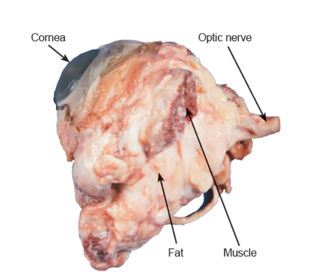
Examine the external characteristics of the eye. Use the image below and identify:
Cornea—at front of the eye; allows light to enter the eye
Optic nerve—at back of the eye; transmits nerve impulses to the brain
Fat
Muscle
Trim the fat and muscle from around the eye using your scissors. Be careful not to cut the optic nerve on the back of the eye. Tip: Use your fingers to pull the fat and muscle away from the eye, then cut it away.
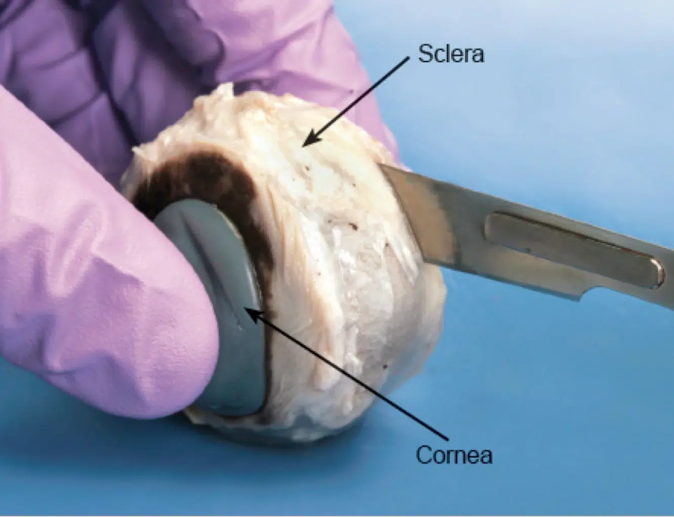
Identify the sclera, or tough outer covering of the eyeball.
Cow Eye Internal Anatomy
Hold the eye between your thumb and forefinger, as shown below. Using scissors or a scalpel, carefully cut the eye in half, separating the front and back of the eye.
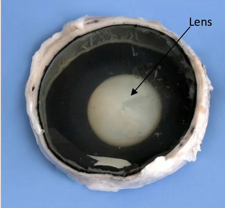
Examine the inside front portion of the eye.
Remove the gelatinous vitreous humor and hard lens. Preservatives make the lens hard and opaque, but in living organisms the lens is clear and flexible.
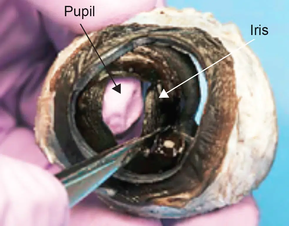
With scissors, cut around the edge of the cornea to expose the iris and pupil.
Use forceps to detach the iris from the sclera and examine the structures.
Examine the back portion of the eye.
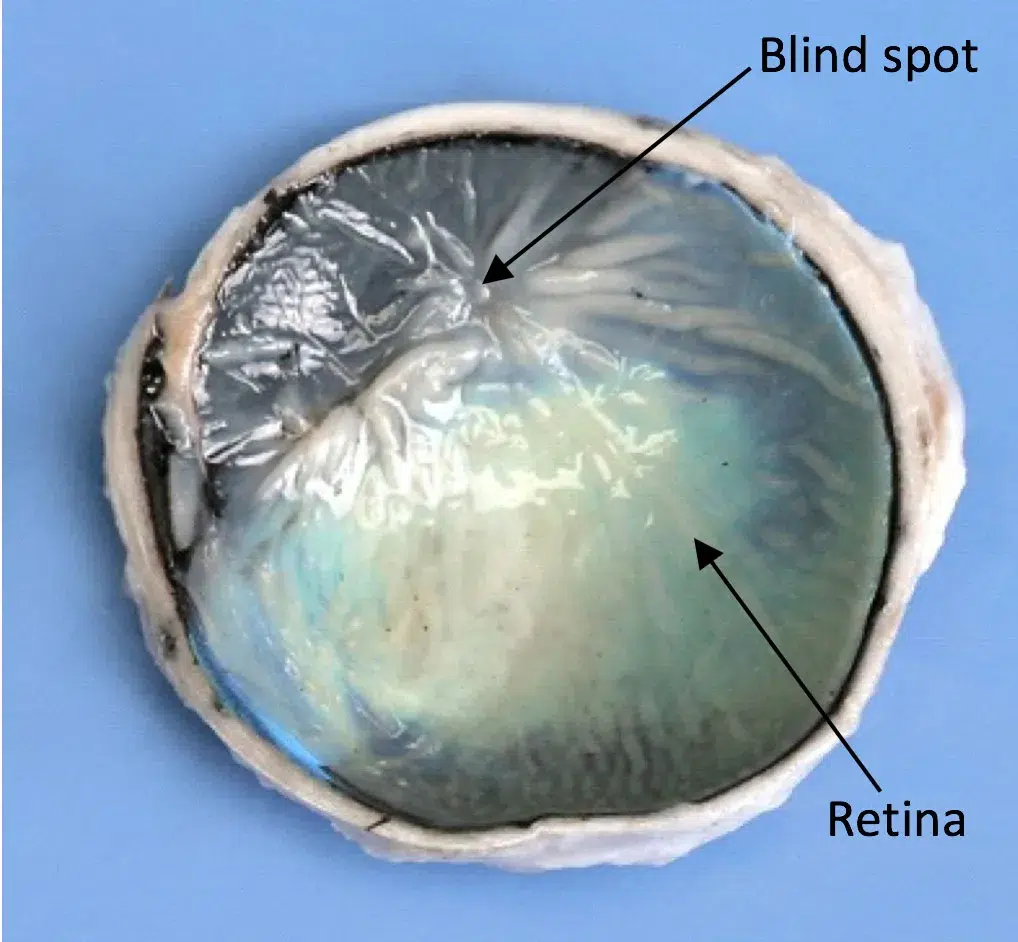
Carefully remove the retina. Notice it connects to the back of the eye at the junction of the optic nerve; this is the blind spot.
Examine the iridescent, reflective layer beneath the retina. This layer is called the tapetum lucidum. Cows and other nocturnal animals have a tapetum lucidum, but humans do not.