CAROLINA DISSECTION GUIDES

Starfish Dissection

Introduction

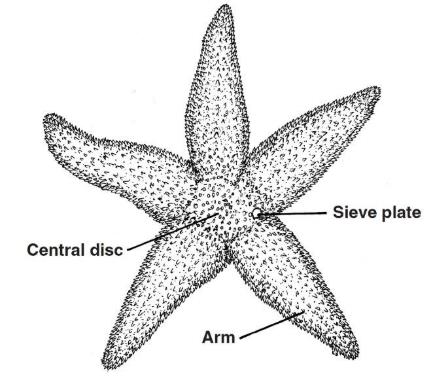
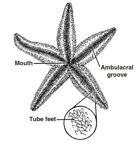
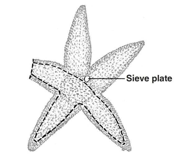
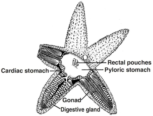
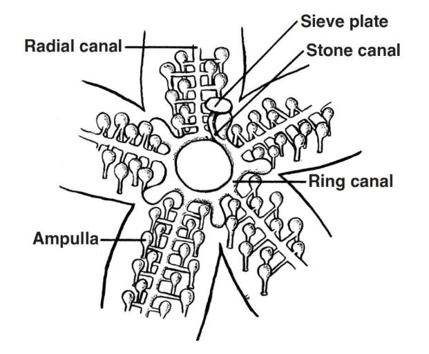
Sea stars, or starfish, are popular dissection specimens for studying the distinctive anatomy of echinoderms. Known for their radial symmetry, tube feet, and water vascular system, these marine invertebrates are an ideal way to connect structure and function in your lab!

Starfish dissection is great for any grade level—from elementary school to college-level comparative anatomy and marine science courses. This activity can also be completed in 1 or 2 lab periods, and students only need dissecting scissors to complete it.

Below is a brief survey of the external and internal anatomy of the starfish. For more detailed dissection instructions and information, check out Carolina® dissection kits.

Time Icon

Suggested Time:

earth science

Ideal Group Size

Safety Supplies

External Sea Star Anatomy

Figure 1. Aboral Surface
  1. Place your sea star on the dissecting tray with its aboral surface facing up, as shown in Figure1.
  2. Examine the radial symmetry of your specimen.  
  3. Observe the protective spines covering this surface. They extend from calcium carbonate plates below the body wall.  
  4. Use Figure 1 to locate the following structures: 
    1. Central disc 
    2. Arms (five) 
    3. Sieve plate, or madreporite 
Figure 2. Oral Surface

5. Turn the sea star over and examine the oral surface (Figure 2). 

6. Locate the following structures:

    1. Mouth 
    2. Ambulacral groove (along each arm) 
    3. Tube feet 

Internal Sea Star Anatomy

  1. Use scissors to cut away the aboral surface from three of the arms, as indicated by the dashed lines on Figure 3. Do not remove the sieve plate.
    Figure 3.

  2. Remove the body wall and locate the paired digestive glands, or pyloric caeca, in each arm (Figure 4).  
  3. Remove the digestive glands from 2 of the arms to reveal the gonads below. 
  4. Identify all the structures in Figure 4. 
    Figure 4.

  5. Remove the pyloric stomach, cardiac stomach, and gonads (from one arm).
  6. Use Figure 5 to locate the following structures of the water vascular system: 
     a. Stone canal
     b. Ring canal
     c. Radial canal (one down each arm)
     d. Ampullae
    Figure 5.
  7. Follow all cleanup and disposal instructions. 

Safety Supplies (Recommended)

About The Author

Newsletter Signup

Get the latest news, free activities, teacher tips, product info, and more delivered to your inbox.