


Anatomy and physiology courses benefit greatly from the use anatomical models and skeletons. Quality models show detailed morphology of organs and complete body systems. Models simplify the identification of anatomical structures, organ positioning within the body cavity, and the relationship between structure and function. Many models can be disassembled, allowing students to explore internal structures. Additionally, they are an excellent reference tool for identifying small and indistinct structures.
Anatomy and physiology is frequently organized and taught by body system. To help you plan and teach, here are our top-selling anatomical body systems models. Click on the model picture or item number for more details.
Integumentary System

Greatly enlarged, this model shows a variety of sensory receptors, as well as the structure of a hair follicle. Layers of the skin can be seen as a cross section and as cutaways at the top of the model. The stratum lucidum is also included to represent thick skin.

This greatly enlarged, block model shows features of epidermal, dermal, and subcutaneous tissues in detail. Sensory and vascular structures, sweat and sebaceous glands, and hair follicles (with and without piloerector muscles) are well represented. Front of hair follicle removes to show internal details.
Skeletal System
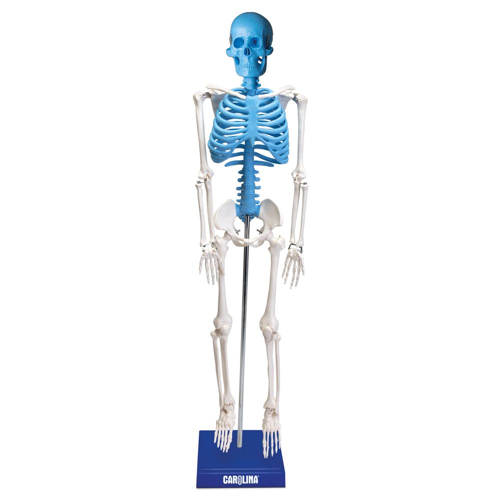
This miniature skeleton stands approximately 85 cm tall and includes a removable skull cap and hinged jaw. There is articulation in the joints. The axial skeleton is in blue, the appendicular skeleton in white, to more easily distinguish the different bones.

Constructed of a durable, unbreakable synthetic material, this skeleton is articulated to show normal posture; the extremities are removable; and the 3-part detachable skull has a cut calvarium, a spring-held lower jaw, and 32 individually inserted teeth.
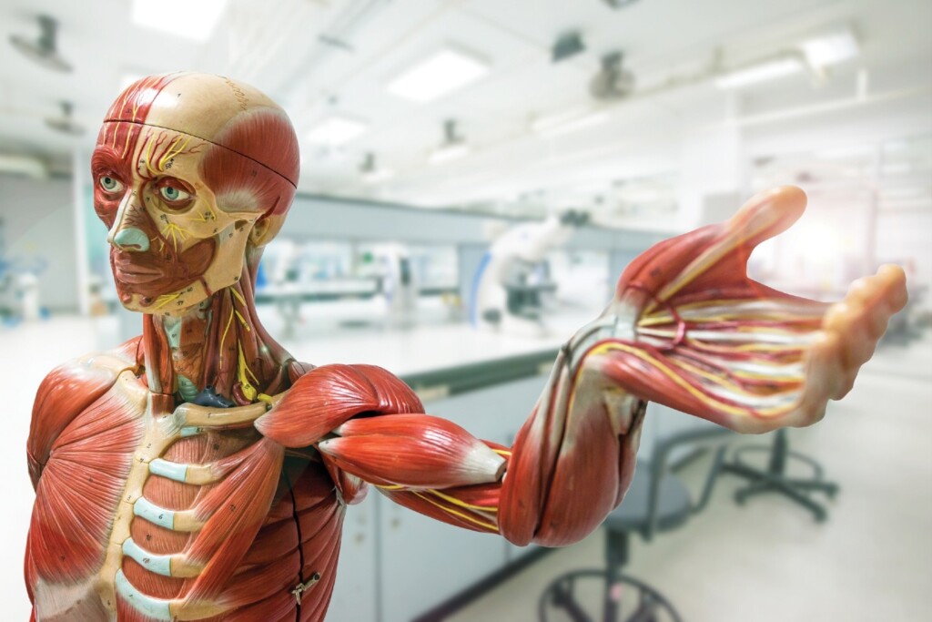
Muscular System
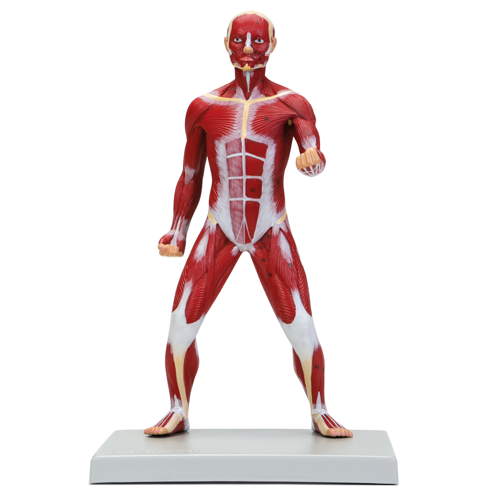
About 1/4× life size and compact, this model is a highly detailed human figure that gives an excellent overview of superficial musculature. All muscles are positioned and sized to correct scale.
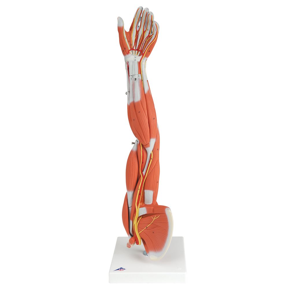
About 3/4× life size, this high-quality muscled arm model illustrates both the superficial and deeper muscles, 5 of which are removable. Tendons, vessels, nerves, and bone components of the left arm and shoulder are shown in great detail.
Nervous System
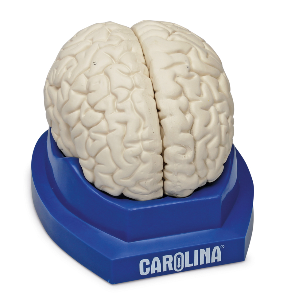
This model is about life size, is dissectible into 3 parts, and identifies the lobes of the brain and 19 internal structures.
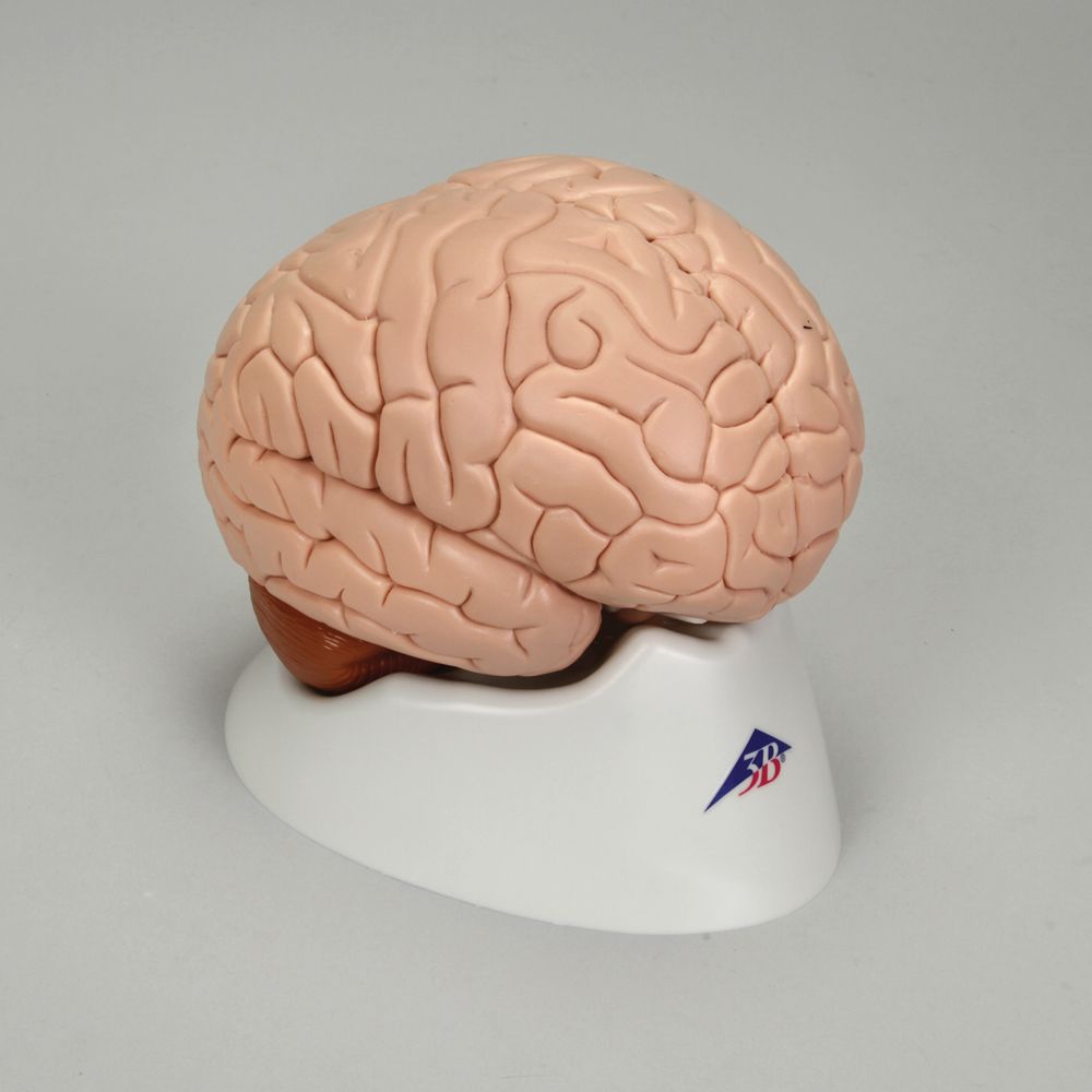
This brain model is life size, dissectible into 2 parts, and shows the median section.
Endocrine System

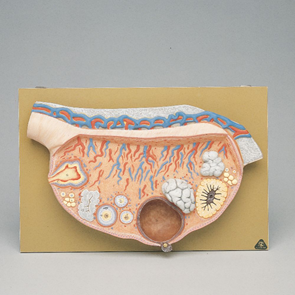
This plaque mount features almost life size, individual representations of pituitary, adrenal, thyroid, and parathyroid glands, plus a testis and a dissected ovary with fallopian tubes and associated structures.
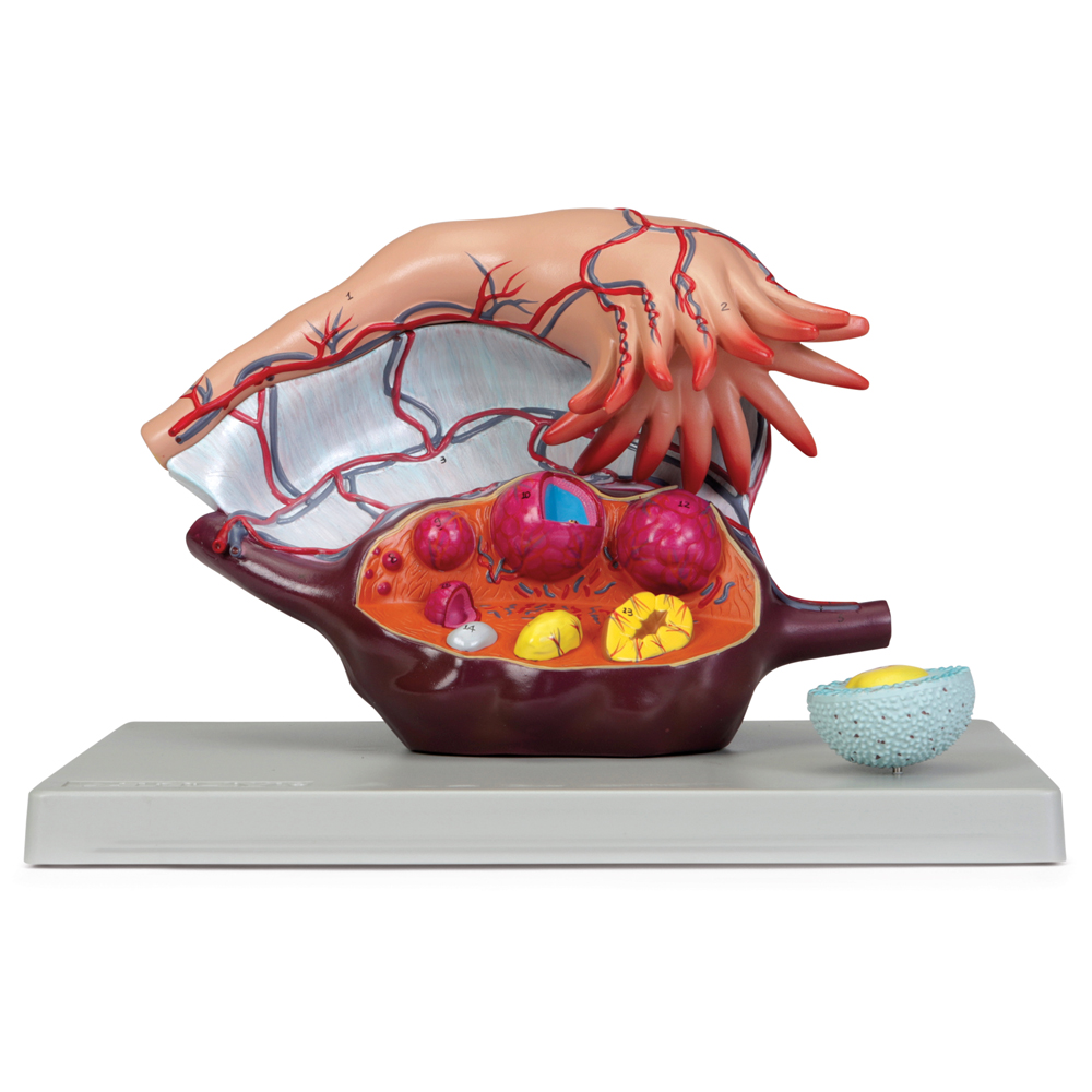
An ovary, almost life size, with removable fallopian tubule shows vascular and suspensory structures. The ovary is sectioned to show follicles at various stages of maturation, from the primary ovarian follicle to the corpus luteum and corpus albicans.
A primary follicle is cross-sectioned to reveal details of the oocyte and zona pellucida.
Cardiovascular System
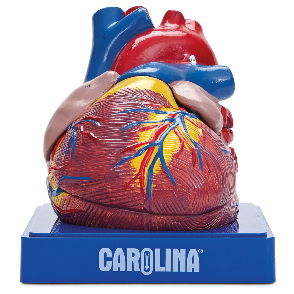
This 2× life size, 3-piece model of the human heart is enlarged for enhanced detail of internal and external structures and identifies 30 structures, including the coronary arteries and veins. The anterior portions of the ventricles and the atria are removeable.
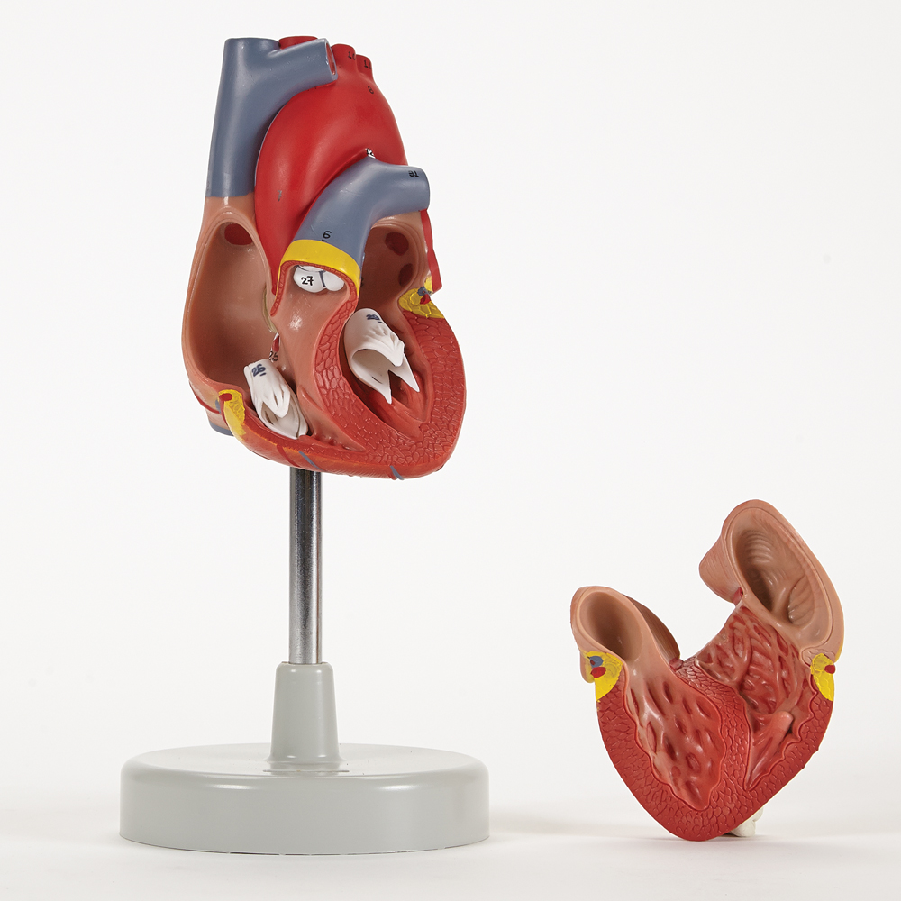
This life size model is dissectible into 2 parts and clearly shows internal and external anatomy including valves, cardiac chambers, and pulmonary and systemic vascular structures.
Lymphatic System
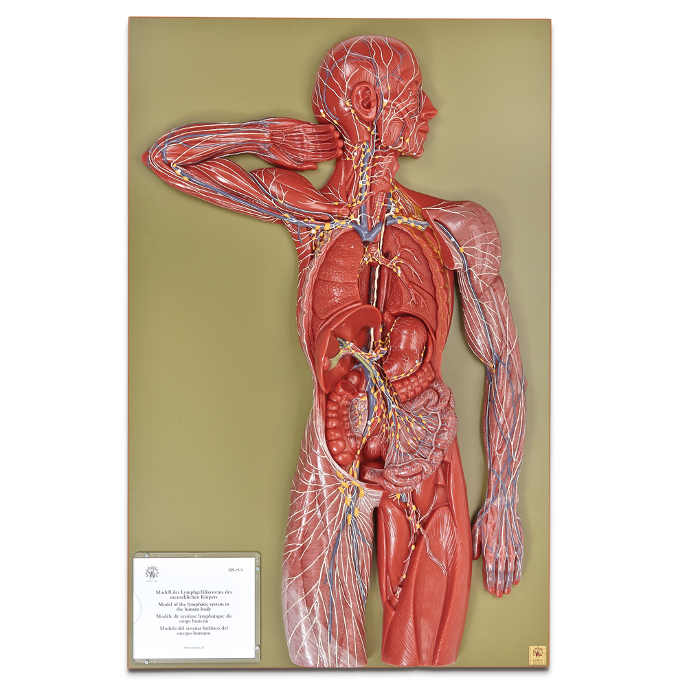
This 2/3× life size, 1-part relief model depicts the human lymphatic system with utmost accuracy and shows lymphatic vessels, ducts, and lymph nodes.

Plaque mount shows erythrocytes, platelets, and 5 types of leukocytes with their characteristic nuclei. Structures are numbered and referenced on the accompanying key. Size, 54 × 38 cm.
Respiratory System
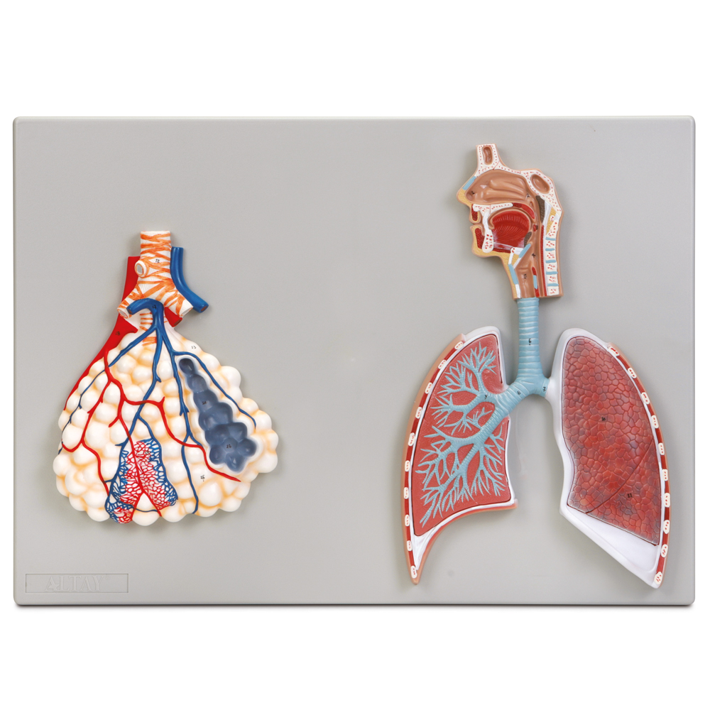
Two-part model shows respiratory airway structures from the nasopharynx to the terminal bronchioles of the right lung. Surface view of left lung shows superior and inferior lobes. Highly magnified representation of pulmonary alveoli shows association with terminal bronchioles and capillary bed.

Realistic depiction of human lung with open dissection to show internal routing of pulmonary arteries, veins, and airways. Pleura and lymph nodes are well represented.
Digestive System
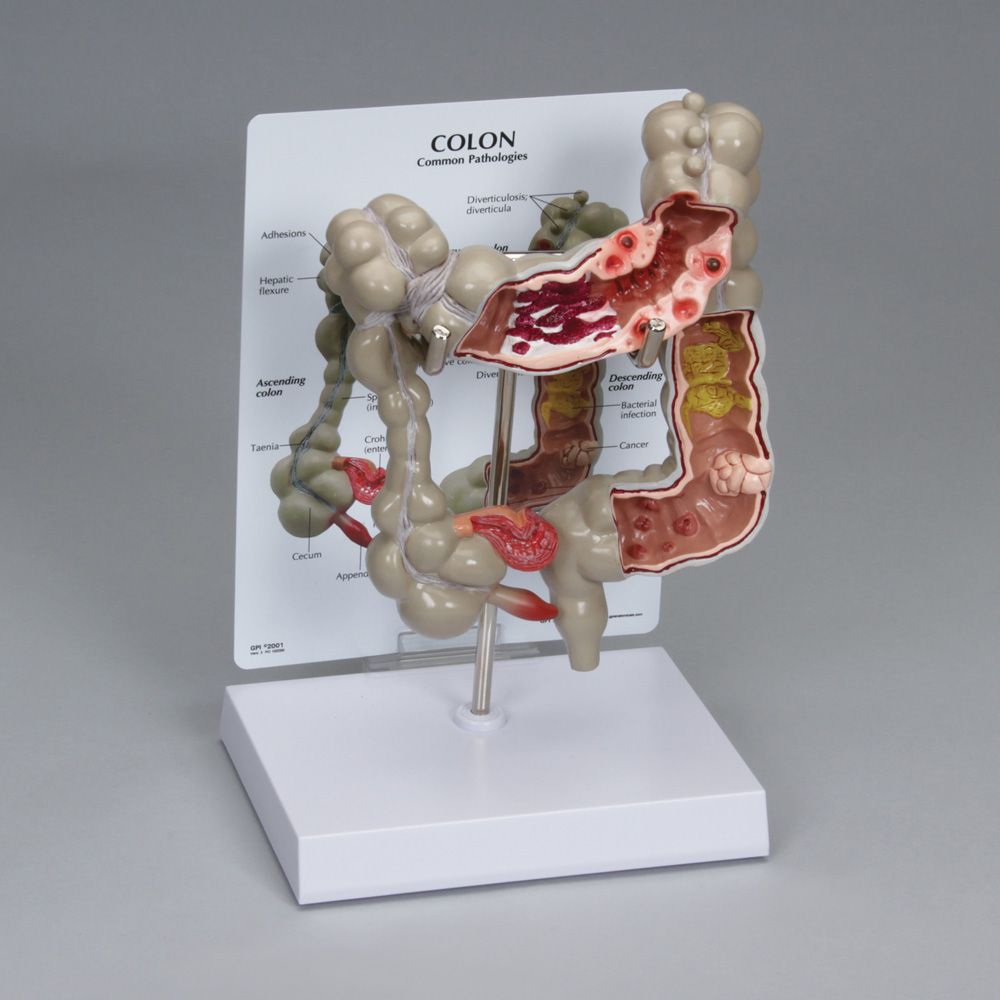
This 1/2× life size model shows common pathologies of the human colon. The ascending, transverse, descending, and sigmoid colon are depicted. Cross sections of the transverse, descending, and sigmoid colon show the following common pathologies: ulcerative colitis, diverticulitis, bacterial infection, cancer, polyps, and more.

This life size digestive system model is dissectible into 3 pieces, and the stomach can be opened.
Urinary System

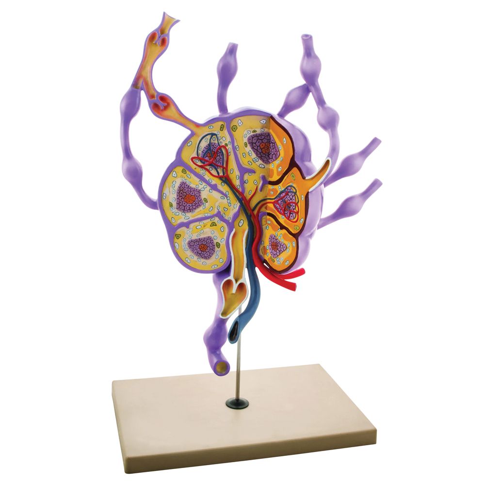
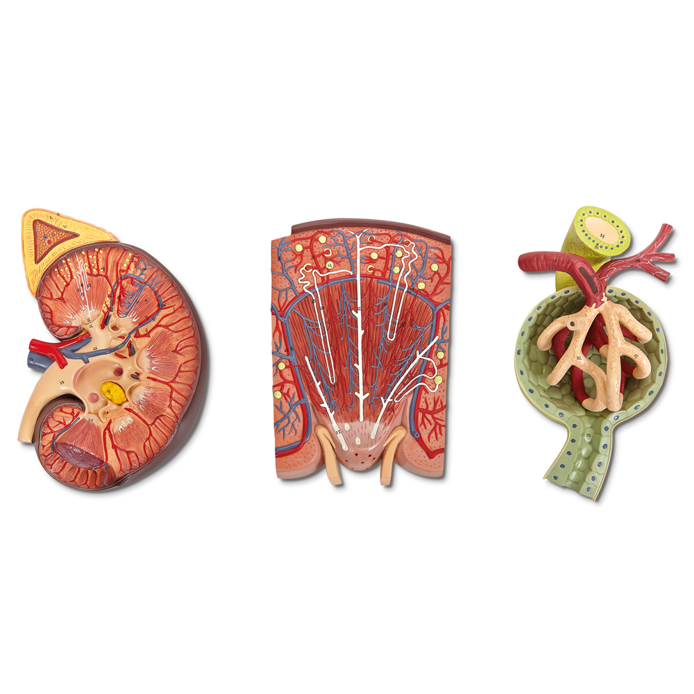
This life size, 2-piece model represents the right human kidney and adrenal gland. The anterior portion is removable to reveal the internal structures of the kidney. The renal artery and vein are shown, as well as the proximal end of the ureter.
This life size, 2-piece model represents the right human kidney and adrenal gland. The anterior portion is removable to reveal the internal structures of the kidney. The renal artery and vein are shown, as well as the proximal end of the ureter.
Reproductive System

About life size female pelvic model allows students to visualize a cross section of a female pelvis including the bladder, uterus, and colon, as well as the structure of the spine and spinal cord present in the pelvis.
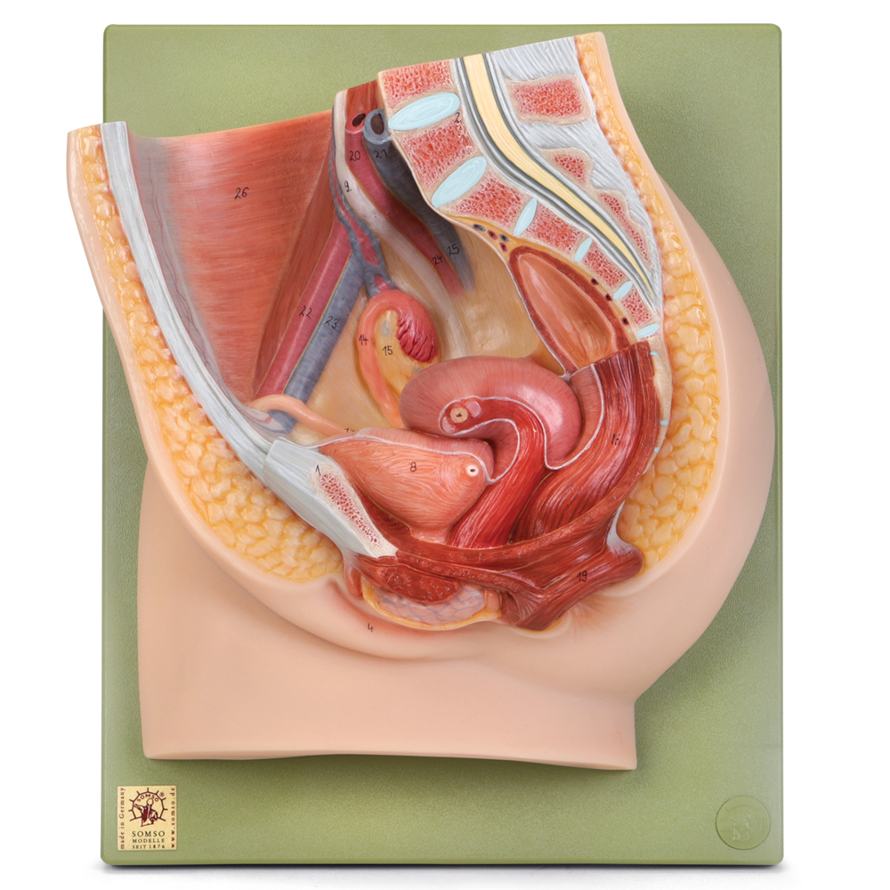
This life size model depicts the right half of the pelvis showing female reproductive organs. The left side of the bladder, uterus, rectum, and ovary are removable.
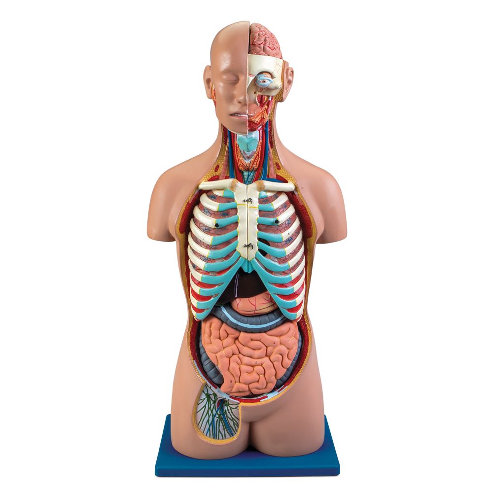
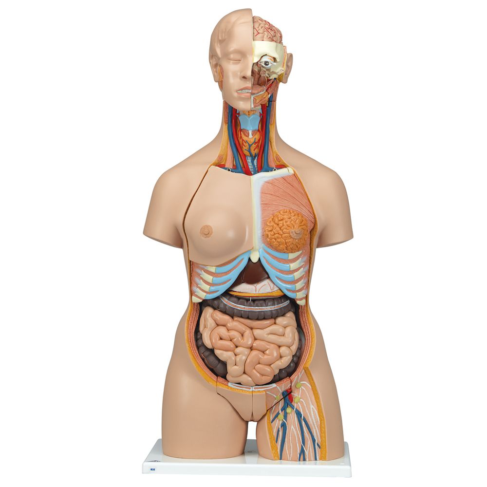
Torso Models

Life size. The Carolina® Human Dual Sex Torso Model with Open Back allows students to study many body systems in one full-sized anatomical model. Students can see the proximity of anatomical structures and view the body as a whole. The 22 removable pieces provide opportunities for more detailed observation of the internal structures of organs such as the heart and stomach. Male and female pelvic inserts are bisected to simplify study of the internal and external reproductive structures.

Life size. Torso features 28 parts, including open back with musculature, vertebral column, paravertebral nerve branches, and removable vertebra with section of spinal cord. One side of model is dissected to reveal skeletal musculature of head and trunk.
Models are a great way to introduce students to body systems and a quick and easy way to review before testing. If you don’t see what you need here, browse our complete line of models. Also, see our free body systems materials and human organs content with infographics and activities to use alongside models. For assistance selecting and caring for your models, we have details here—and don’t forget specialized cleaning solutions to ensure your models remain like new.