Fern Life Cycle

Published: April 1972 | Updated: May 2026

Ferns and their allies, like the horsetail, have thrived for over 300 million years and are one of the most ancient and diverse groups of vascular plants on Earth. Today, botanists recognize about 10,500 extant species, but the number may be higher due to ongoing discoveries in tropical regions. Their deep evolutionary roots, unique structural features, and ecological adaptability make them a fascinating subject of botanical study. They draw attention from everyday gardeners because they are widely distributed and many are easy to grow. Because of their ancient lineage and their central position in the plant kingdom’s evolutionary history, ferns draw plenty of interest.

History and Evolution of Ferns

Ferns first appear in the fossil record during the Middle Devonian Period, about 383–393 million years ago—and may have originated even earlier. These early ferns included now-extinct lineages such as the Rhacophytales and ancient tree ferns like Pseudosporochnales. Ferns expanded extensively during the Carboniferous Period (358 to 298 million years ago), an era often referred to as the “age of ferns.” The Carboniferous Period was a lush, green time of fern-dominated forests that contributed significantly to Earth’s current coal deposits.

Both leptosporangiate and eusporangiate ferns in the crown group, likely originated in the late Silurian period (around 423 million years ago), with major diversification occurring during the Cretaceous Period. Despite competition from seed plants and flowering plants, ferns persisted and infiltrated many habitats, becoming the second-most diverse group of vascular plants today.

Structures and Morphology

Ferns are vascular plants, having true roots, stems, and complex leaves (megaphylls) which distinguish them from non-vascular plants like mosses. A defining feature of the fern is the mesarch protoxylem, a developmental trait uniting all ferns despite their diversity. Most species of ferns belong to the leptosporangiate ferns, which produce spores in delicate, single-celled-origin sporangia often clustered into sori.

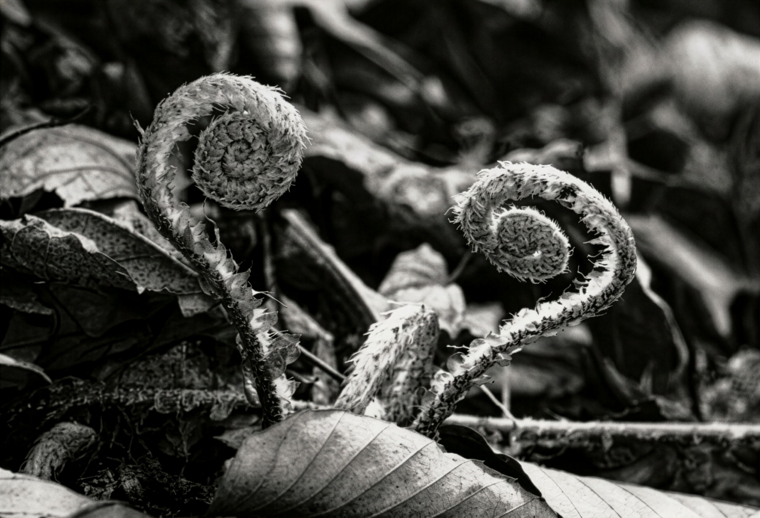
The fern life cycle alternates between a dominant sporophyte stage and an independent gametophyte stage, with reproduction happening via spores rather than seeds. New leaves typically emerge as tightly coiled fiddleheads, which unfurl into fronds.

Ferns display extensive morphological variation. They may be:

  • Terrestrial, rooted in soil or moss,

  • Epiphytic, growing on tree trunks or branches,

  • Aquatic, either floating or submerged.

Their size ranges from 1 cm tall filmy ferns to tree ferns reaching 25 meters, illustrating exceptional structural diversity.

Sporophyte

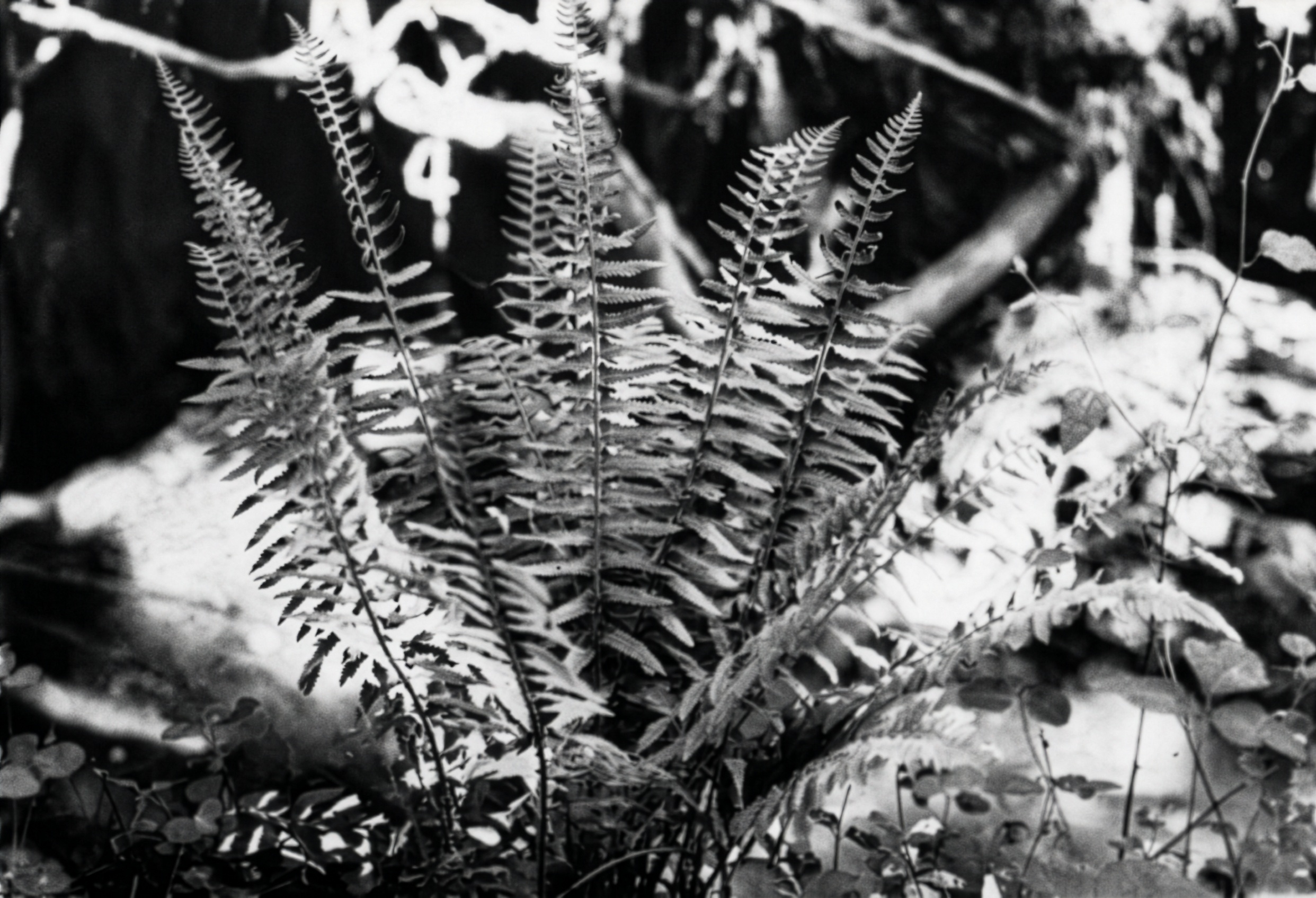
The familiar fern plant is a sporophyte (Fig. 1), and is most often found in shady woods. The fern sporophyte has true roots, stems, and leaves, and has a well-developed vascular system.

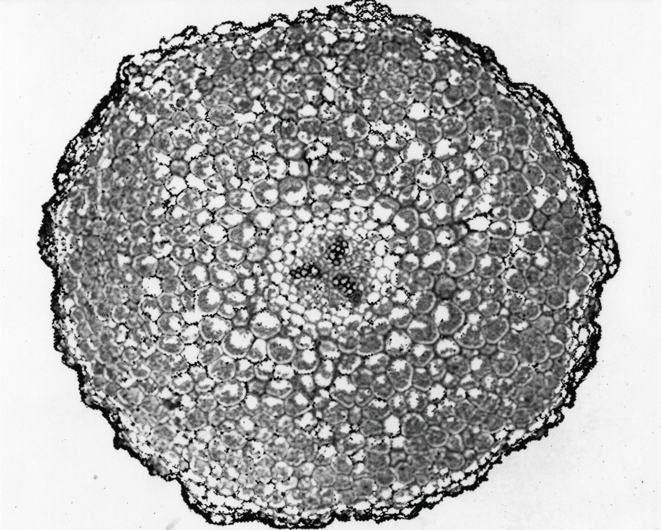
The roots in the mature fern plant are adventitious and arise from the stem (rhizome) between the leaf bases. The adventitious roots of ferns typically show a radial arrangement of xylem and phloem (Fig. 3). The number of xylem strands may vary but there is always an alternation of xylem and phloem strands. This arrangement is common to the roots of the higher plants.


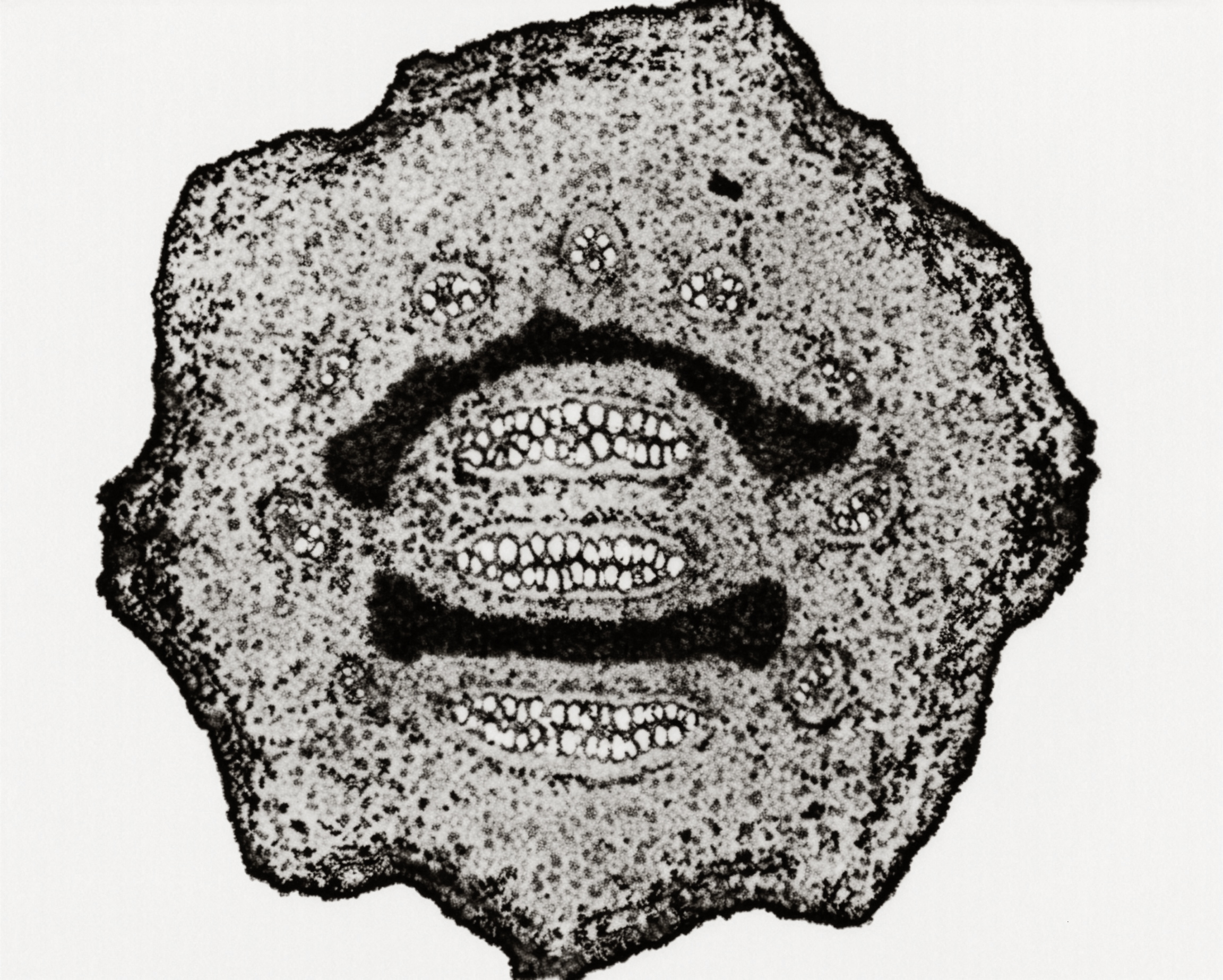
The stem is usually a horizontal, underground rhizome. There’s great diversity in the structure of the vascular systems of the different fern genera. A common vascular arrangement is the dissected siphonostele found in bracken fern (Fig. 4). Here the small vascular strands are arranged in a circle.

There is a wide range in leaf form and size among the ferns. Most species have compound leaves, often called fern fronds (Fig. 1), with leaflets called pinnules. The leaf bud (fiddlehead, Fig. 2) uncoils from the base to the tip. The leaf continues to grow at the tip until it reaches maturity.

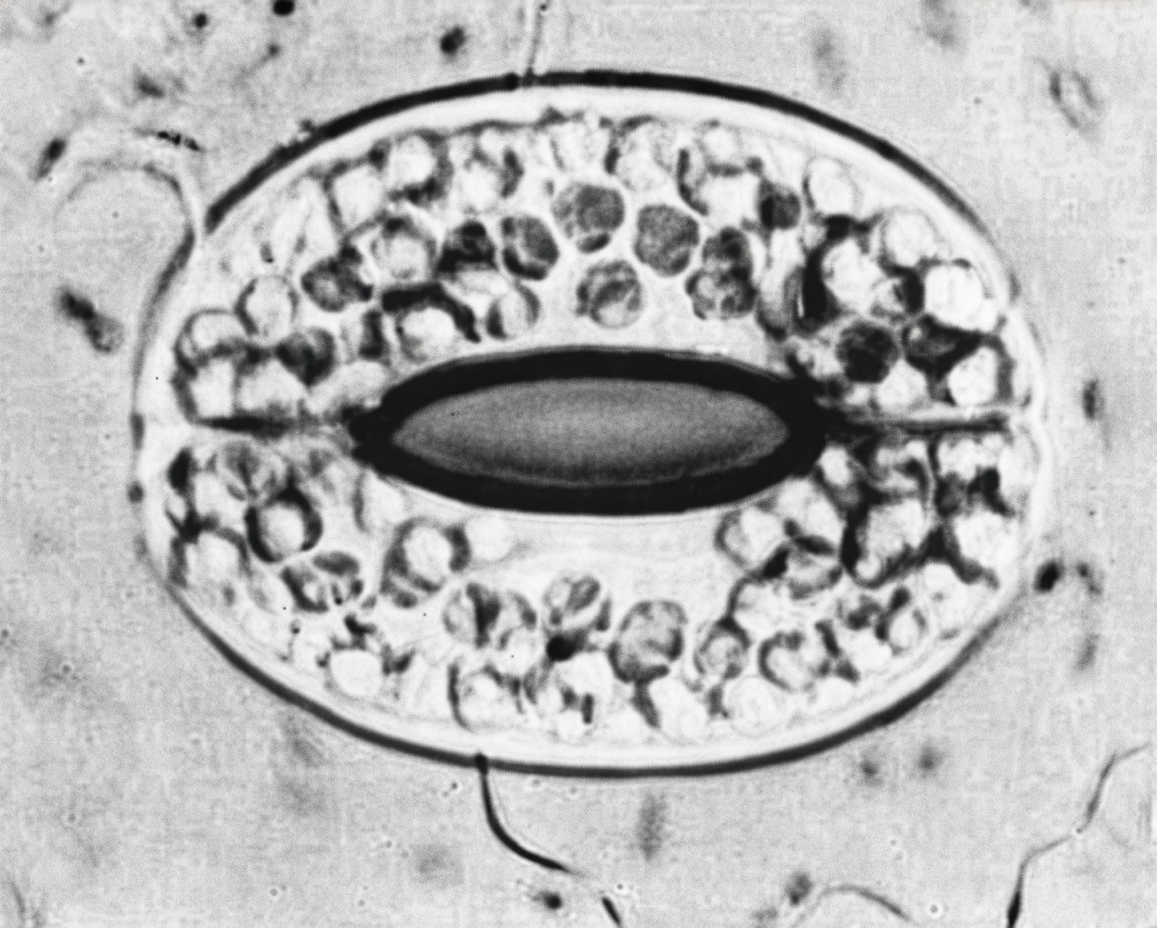
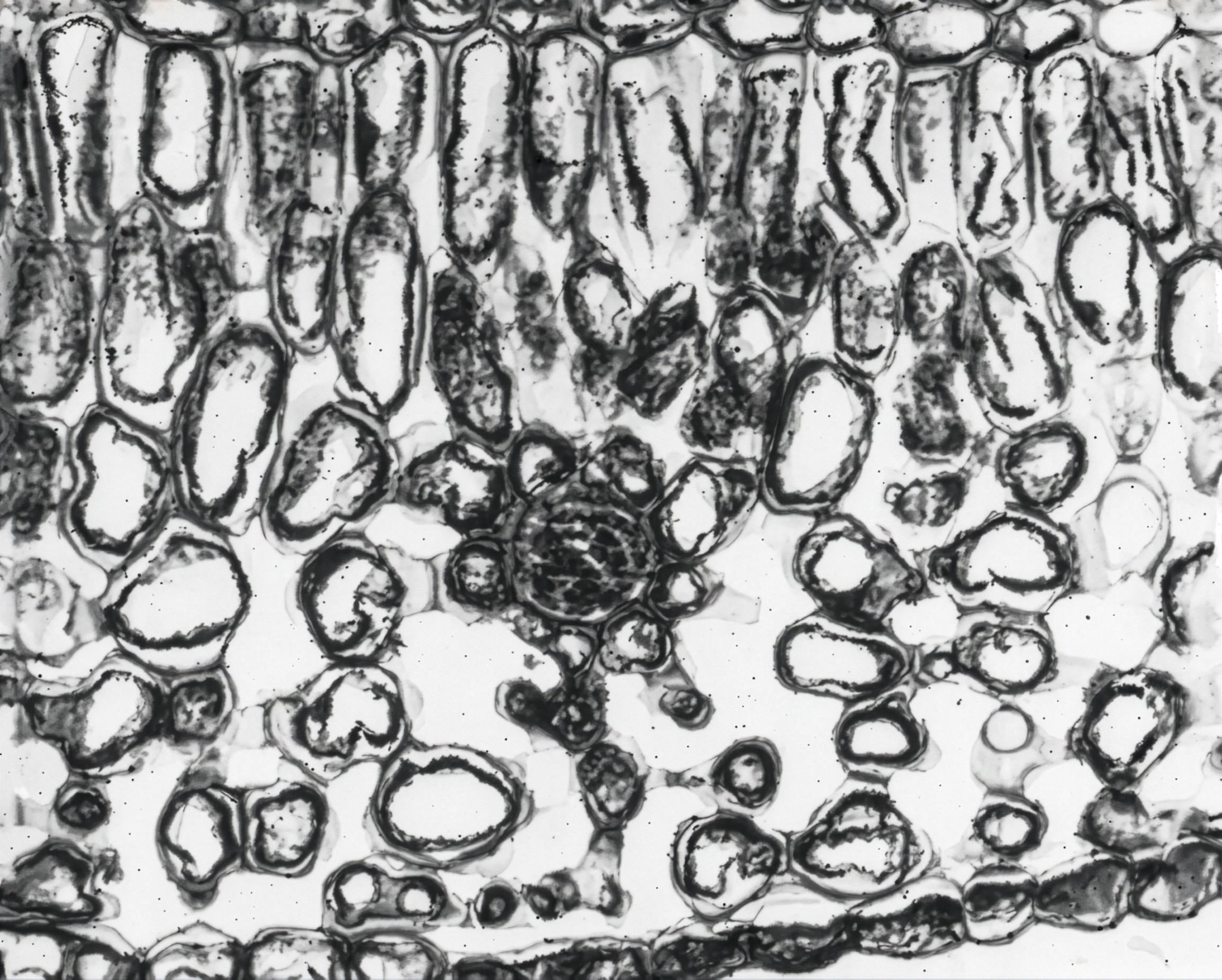
The outer layer of leaf cells is the epidermis, with stomata usually found in the lower epidermis of the pinnule (Fig. 5). The internal structure of the leaf (Fig. 6) is similar to that found in the leaf of seed plants. Just below the upper epidermis is a group of elongate cells making up the palisade tissue. Beneath the palisade cells are spongy mesophyll and vascular tissues.


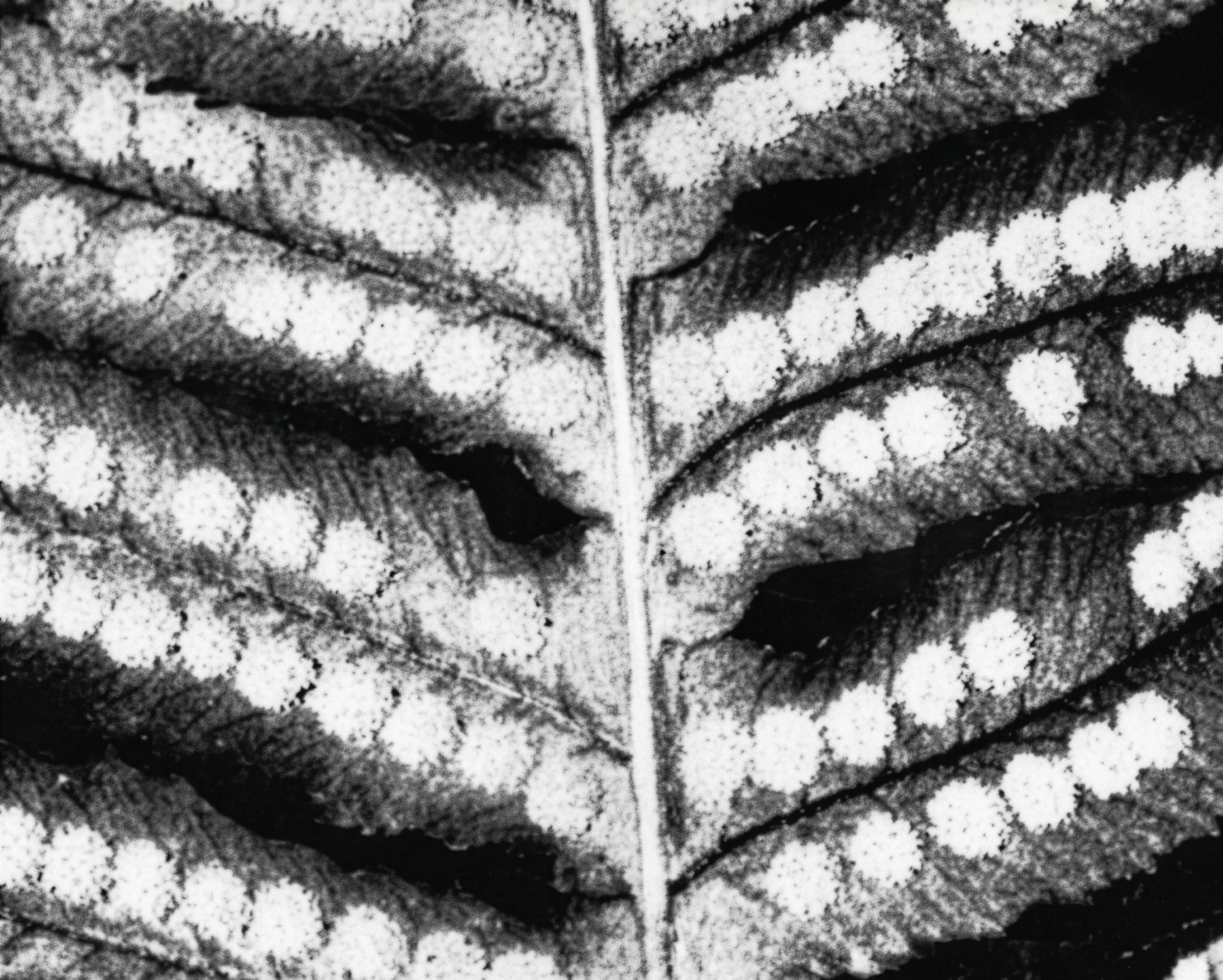
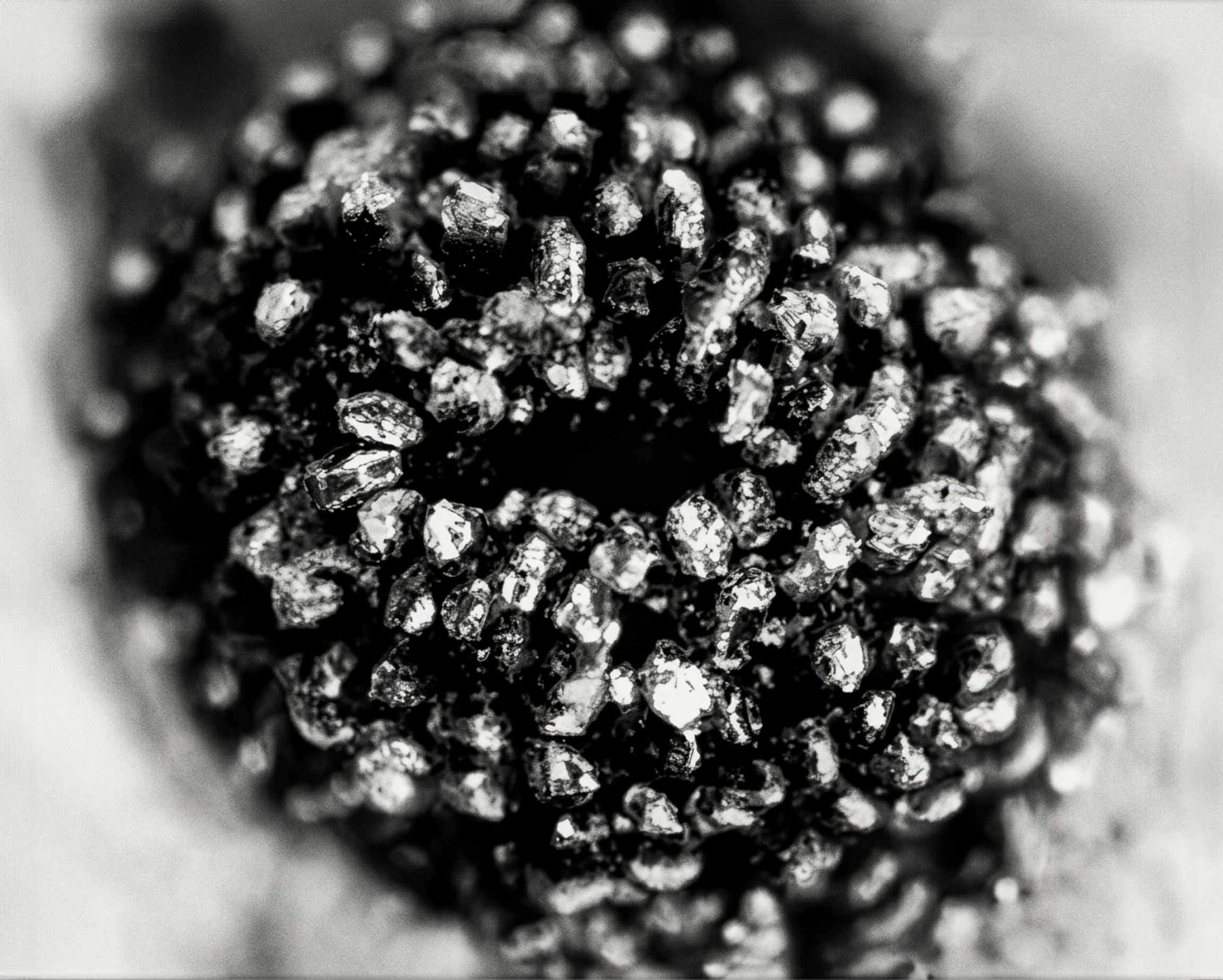
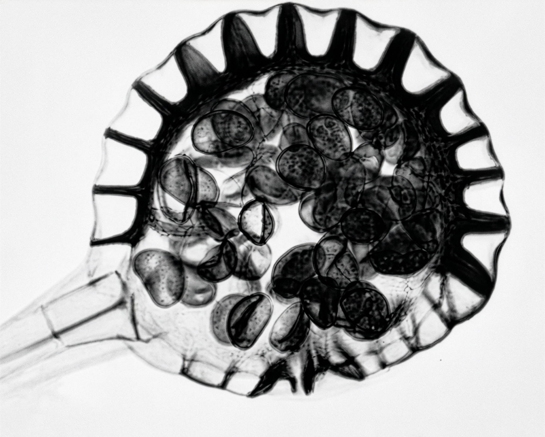
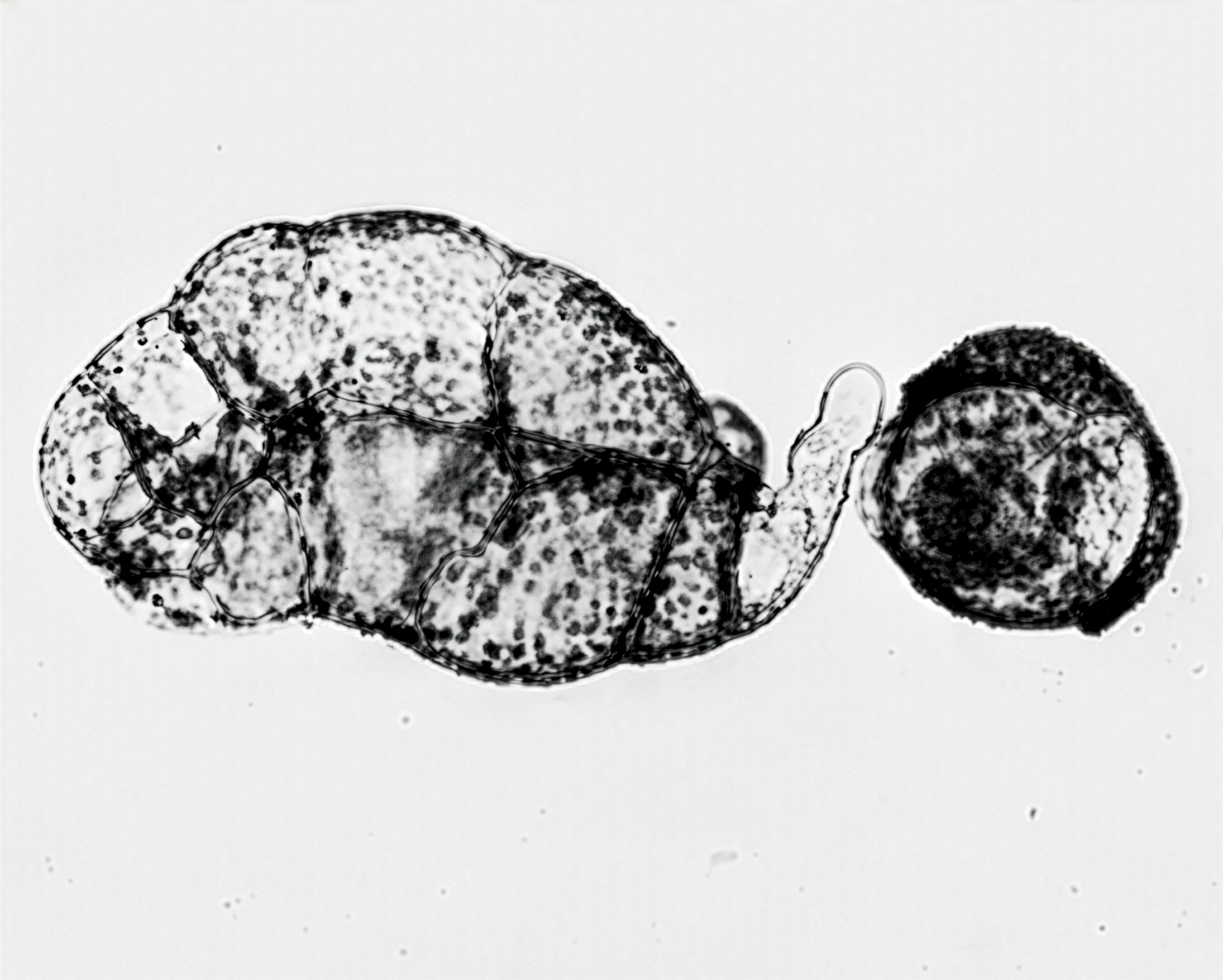
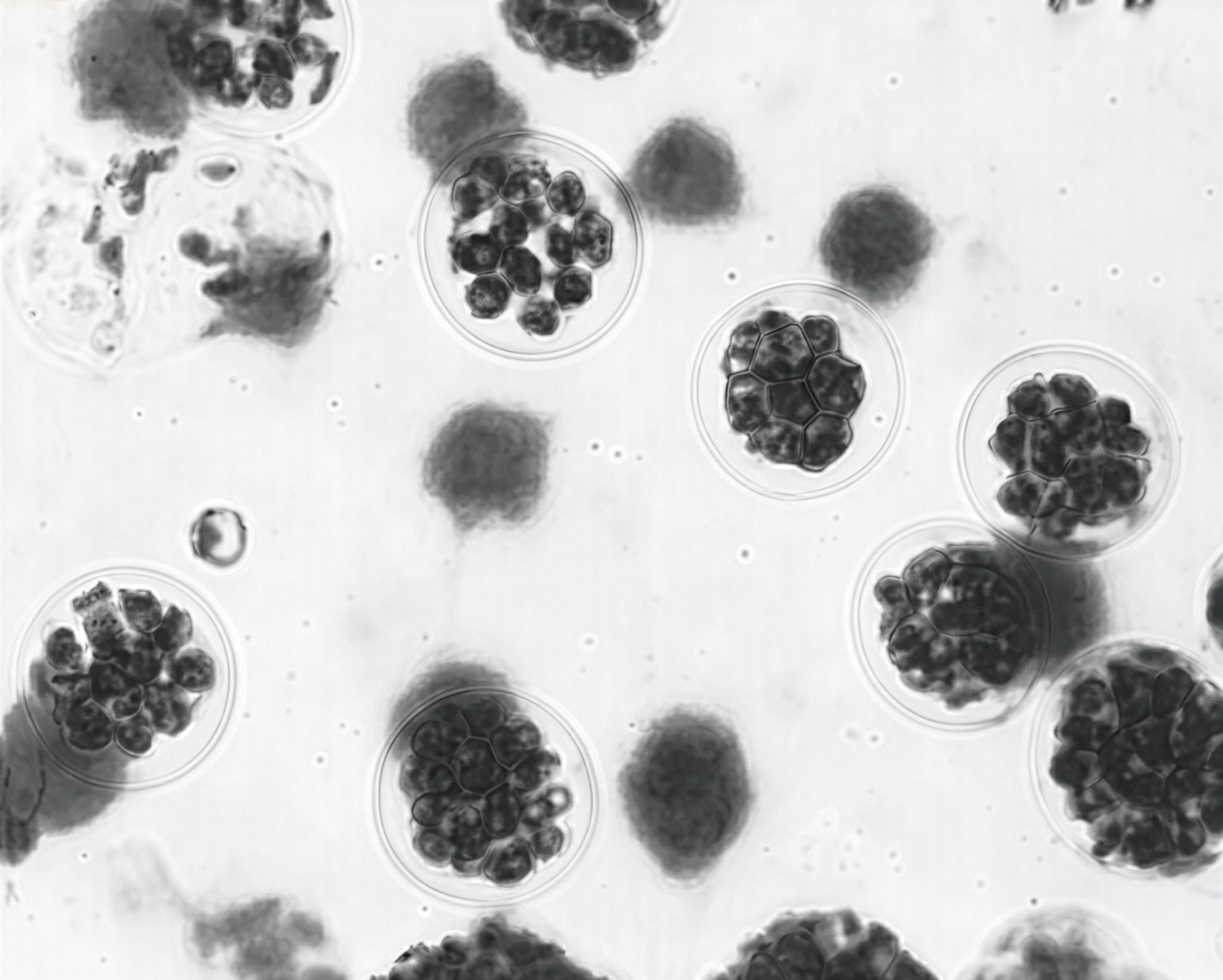
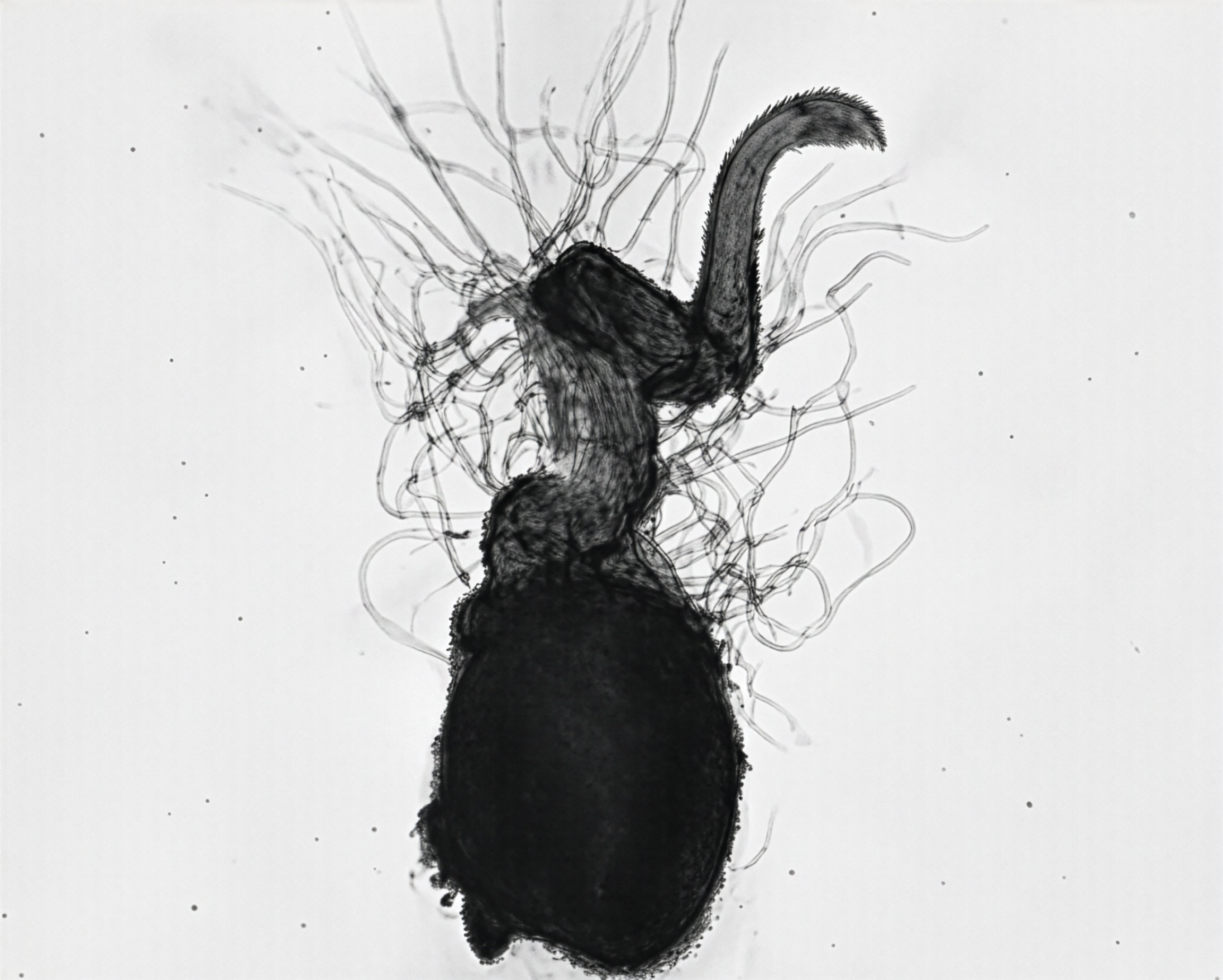
In many ferns the sexual phase begins on the underside of the vegetative leaves in specialized structures called son (Fig. 7). Each sorus is a cluster of smaller structures known as sporangia (Fig. 8). Each sporangium usually consists of a stalk and a capsule (Fig. 9). The capsule wall is modified to form an annulus, a group of thickened cells and lip cells.

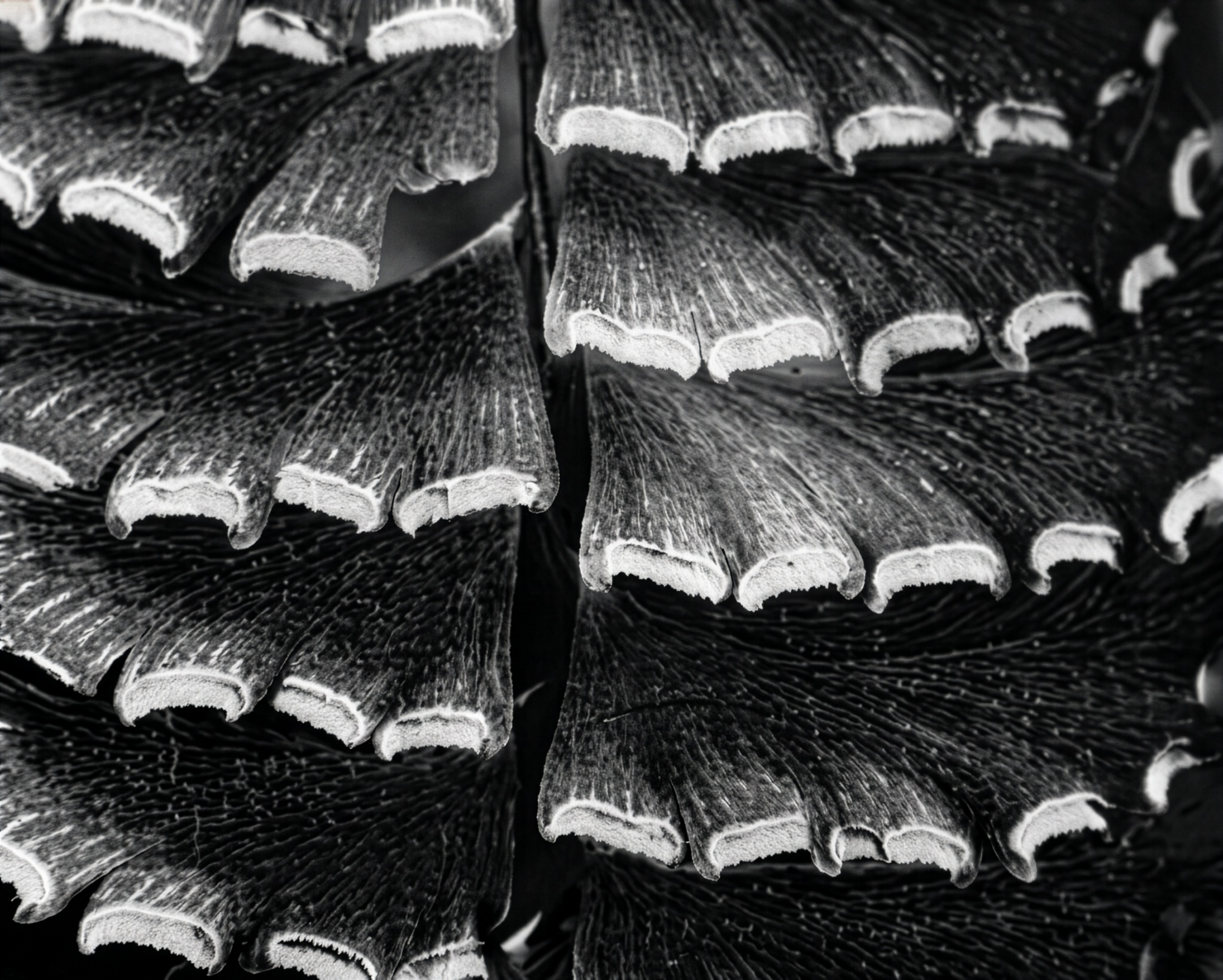
The sorus may be covered by an umbrellalike structure called an indusium (Fig. 10). In most cases, the indusium is a special outgrowth of the leaf, and as the sporangia mature, they may protrude from beneath the indusium. When the covering is formed by the inrolled leaf margin, it’s called a false indusium (Fig. 11).

FIGURE 1 Sporophyte of Christmas fern.
FIGURE 2 Fiddleheads of cinnamon fern.

Gametophyte

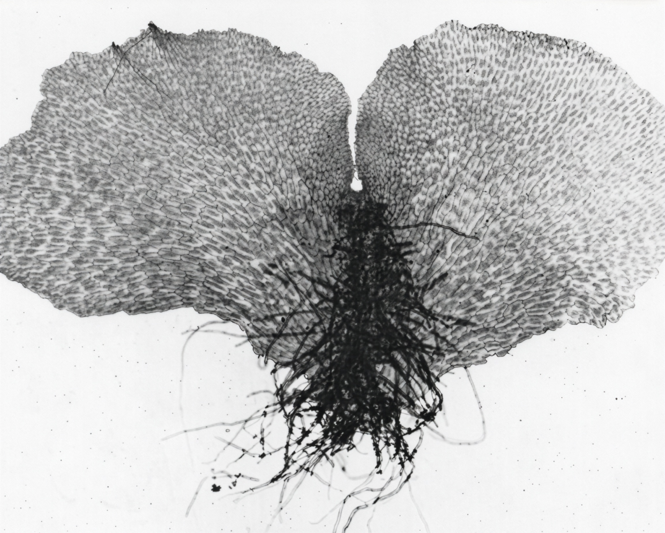
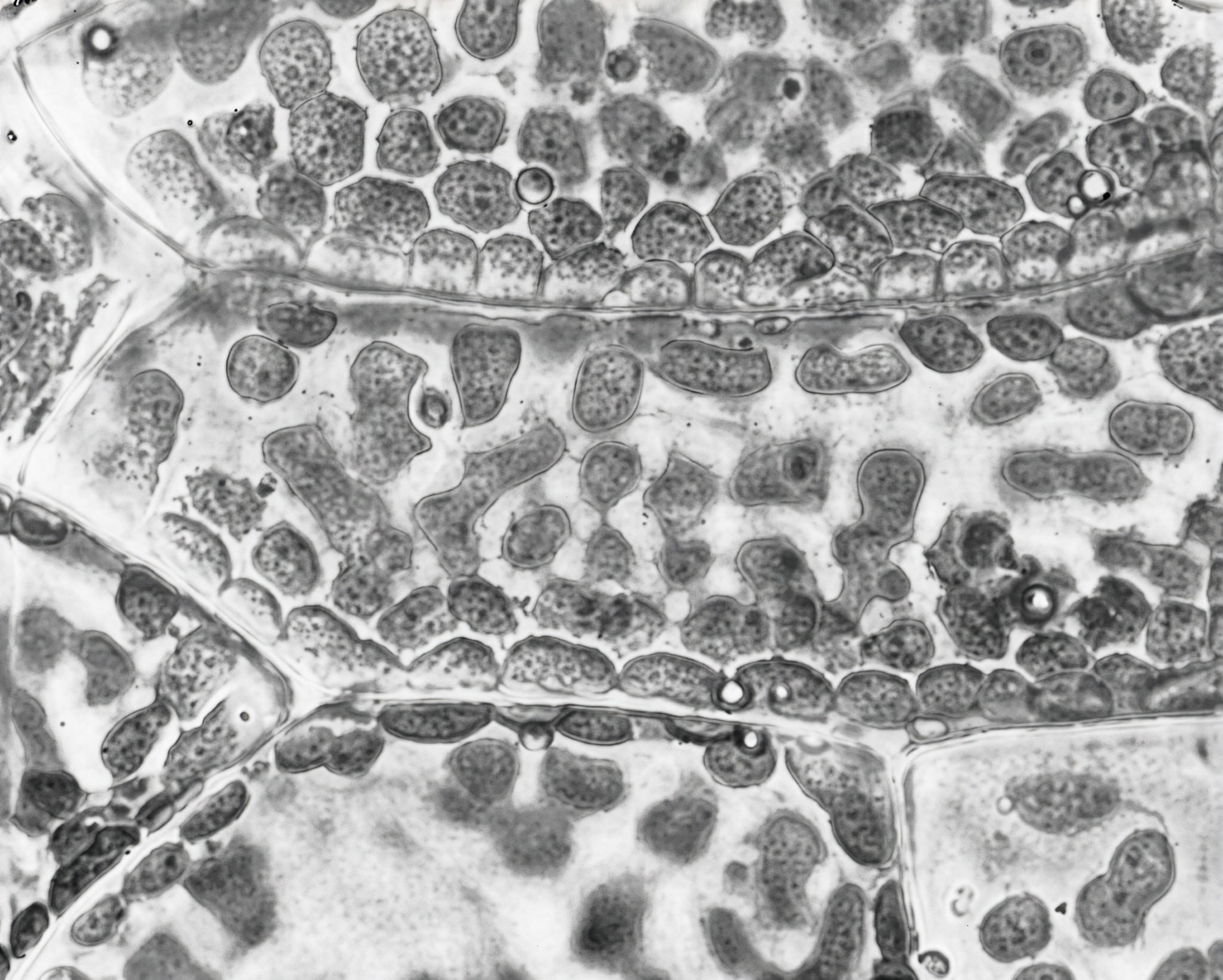
A fern spore germinates and produces a gametophyte, a flat plate of cells called a prothallus (Fig. 12). The resulting growth is mostly two-dimensional and produces a thin ribbonlike or heart-shaped prothallium with several rhizoids (Fig. 13). The prothallial cells are densely packed with chloroplasts (Fig. 14) which give the young gametophyte a rich green color. 

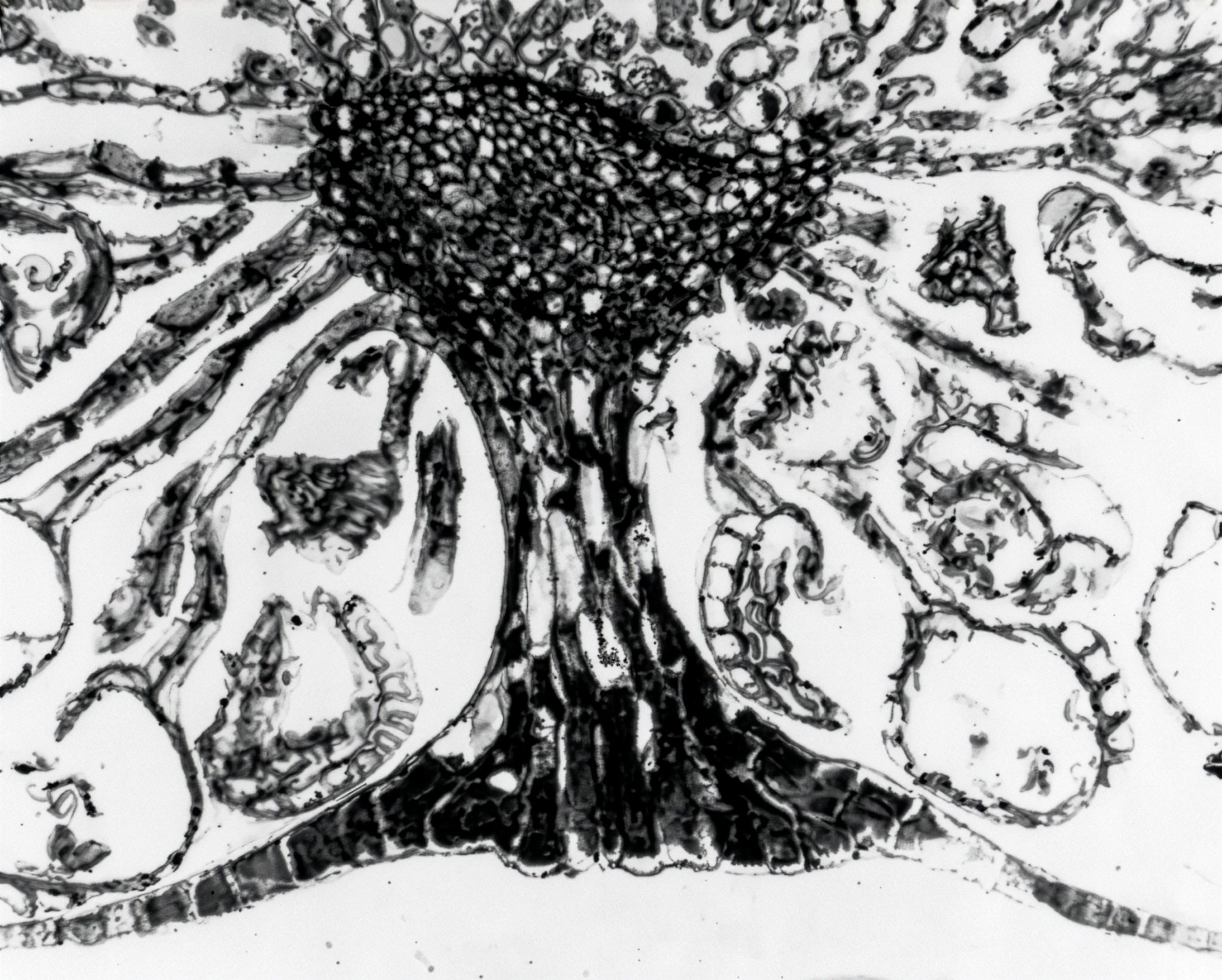
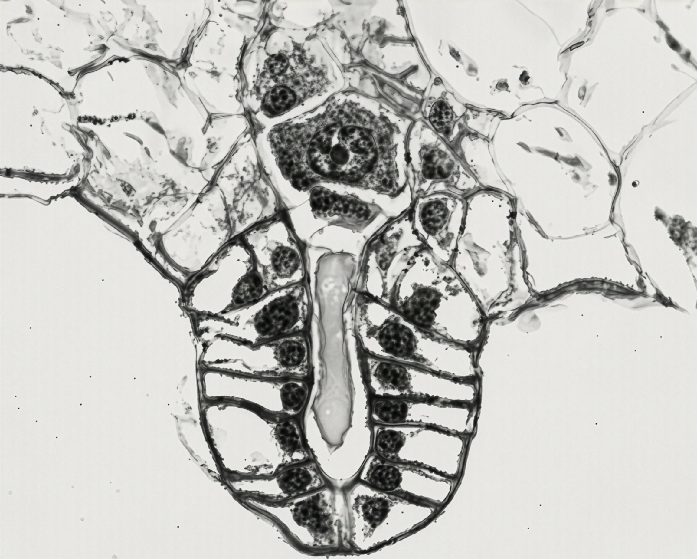
A typical heart-shaped prothallium (Fig. 13) has numerous rhizoids and reproductive structures on the lower surface. Archegonia, usually few in number, normally develop near the apical area. An archegonium consists of a sunken venter, a short neck, two neck canal cells, a ventral canal cell, and an egg (Fig. 15).


Antheridia generally develop near the rhizoidal area of the prothallium (Fig. 16). In liquid culture, archegonia and antheridia usually appear on separate prothallia. Spermatogenous cells form in the developing antheridium and differentiate into corkscrew-shaped multiflagellate sperm (Fig. 17). A sperm may enter the archegonium through the neck canal and fuse with the egg (Fig. 18), these being the gametes. The fertilized egg (zygote), which is diploid, is the beginning of the sporophytic generation.

Sporophyte

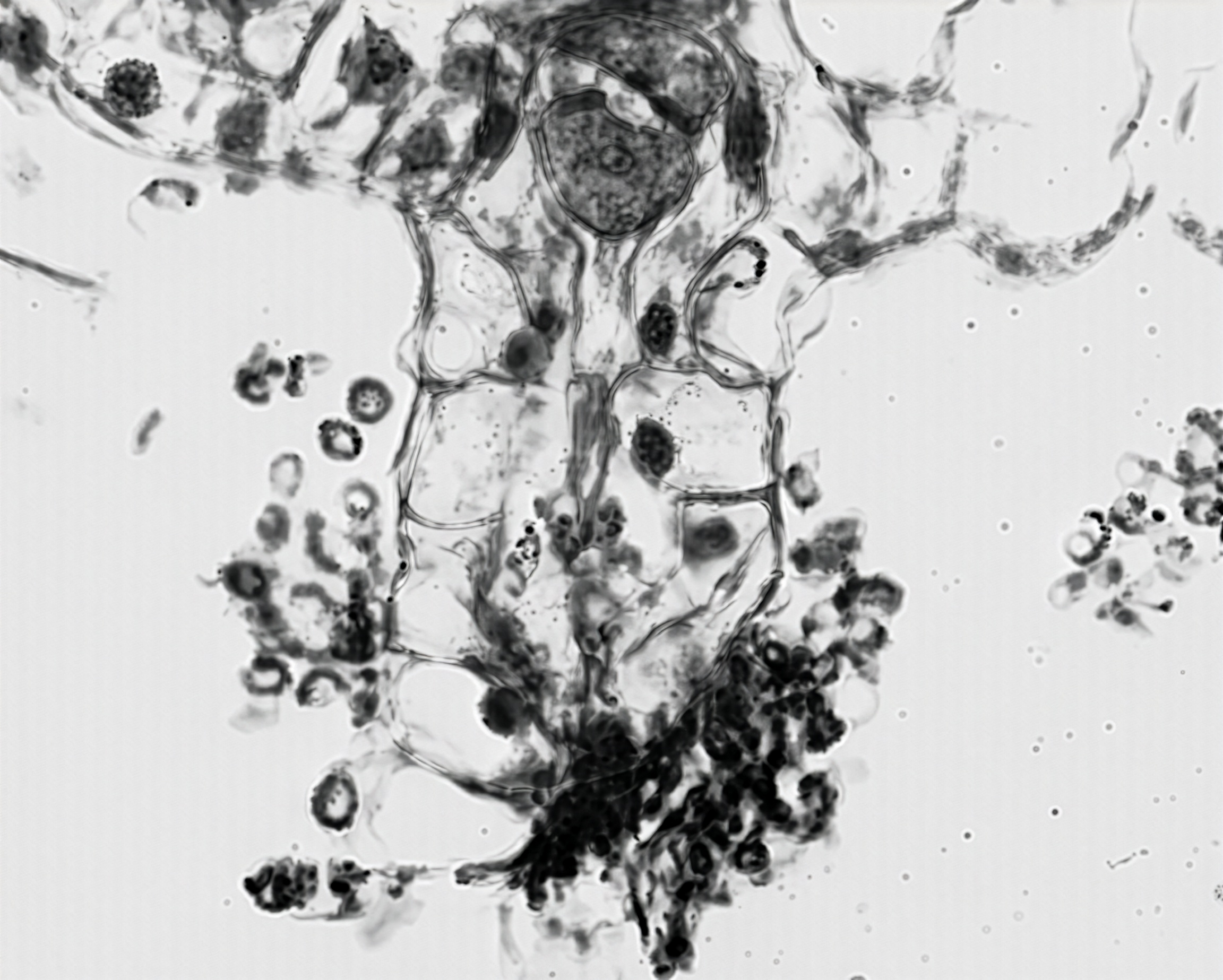
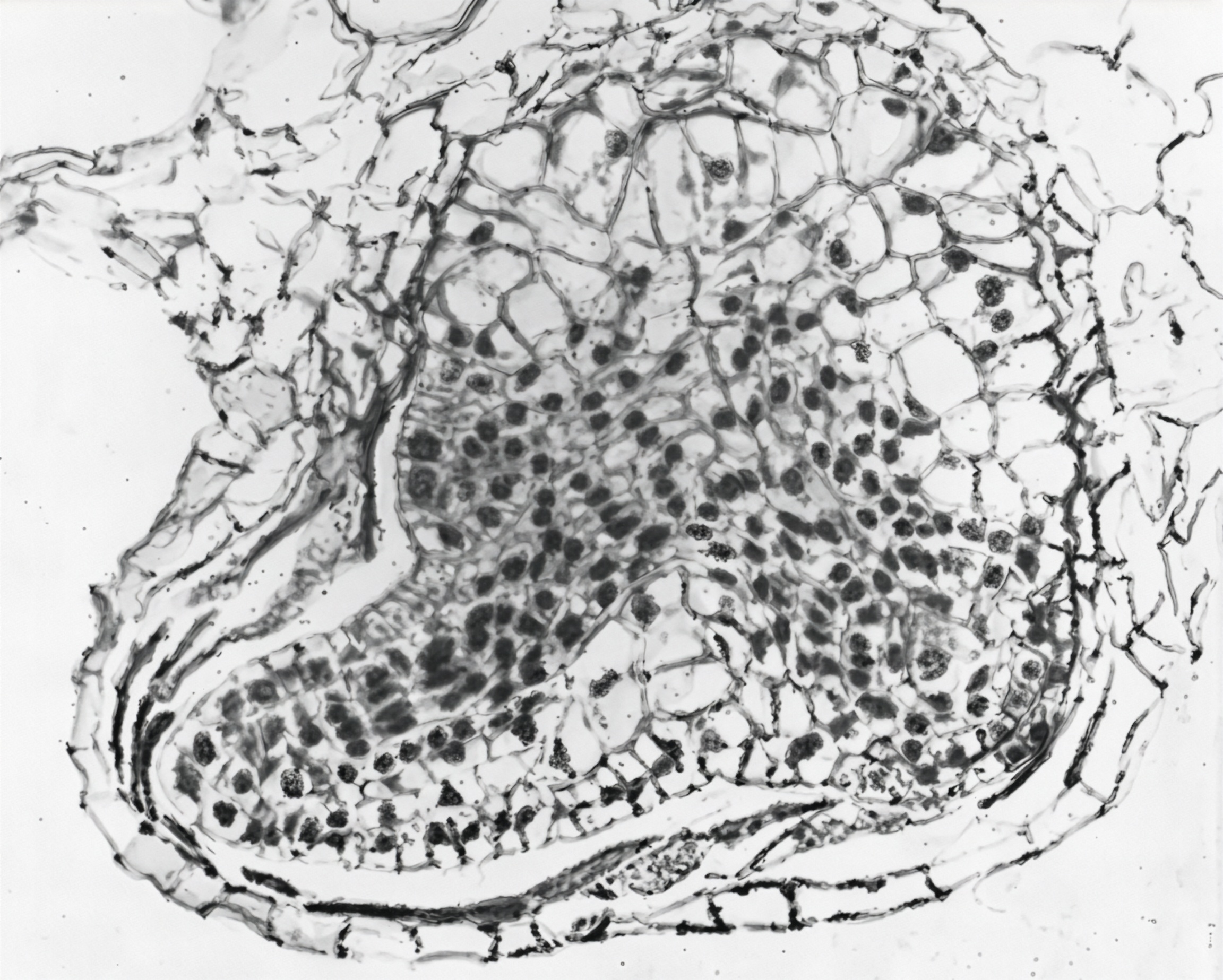
In fern reproduction, the zygote stays in the archegonial chamber and develops into an embryo (Fig. 19). The embryo has four growth areas. These areas develop into a foot, a primary leaf, a stem, and a root. The young sporophyte is embedded in the gametophyte and absorbs nourishment from it through the foot.

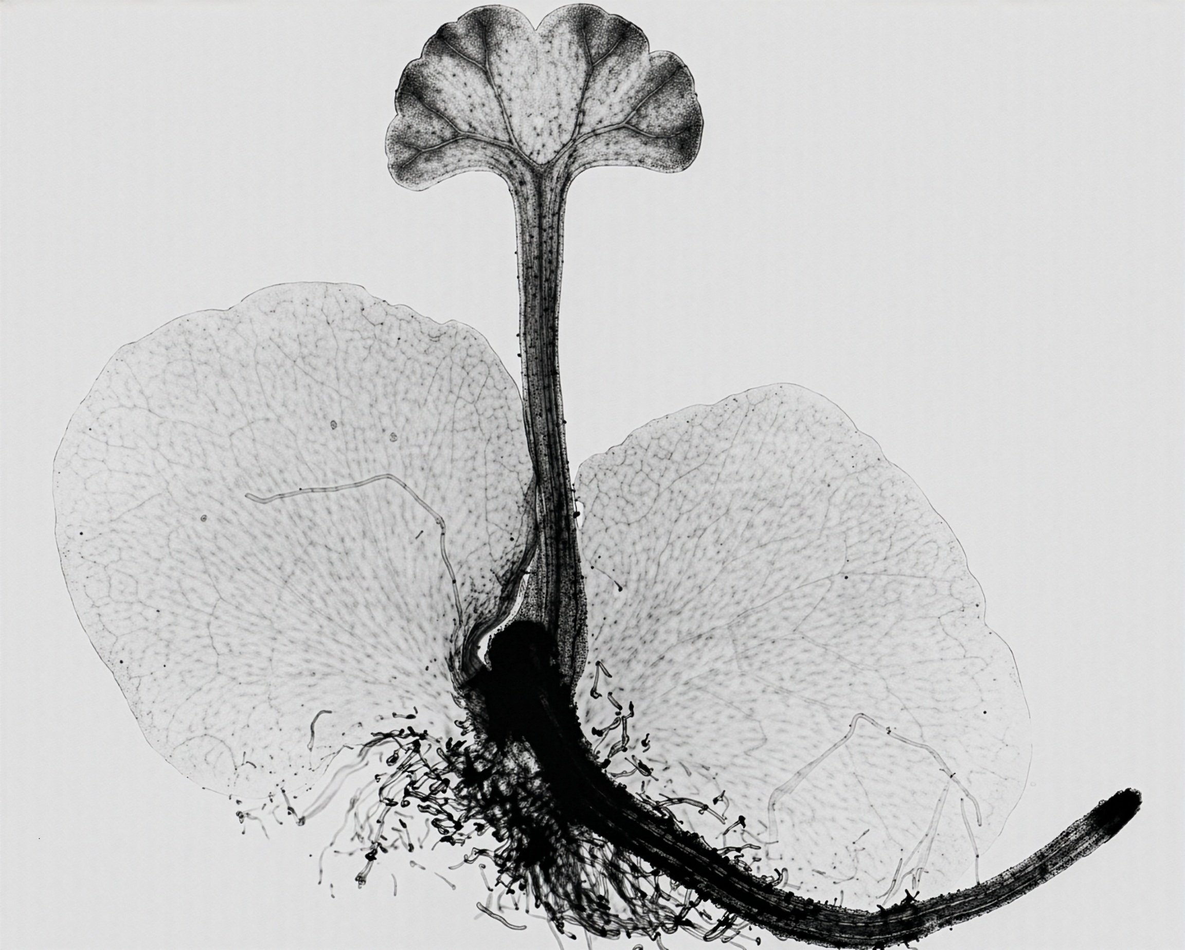
As the young sporophyte develops, it breaks through the archegonial wall. This is followed by growth and development of the primary leaf and root (Fig. 20). Ultimately, the stem grows into the soil and produces leaves and adventitious roots. The gametophyte, primary root, and primary leaf die, and the dominant sporophyte of mature plants is established. With that, the life cycle of a fern, characterized by an alternation of generations, is completed. Images of adult ferns and embryos are below.

FIGURE 3 Root of Botrychium, c.s.
FIGURE 4 Rhizome of Pteridium, c.s.
FIGURE 5 Fern stomata.
FIGURE 6 Christmas fern leaf, c.s.
FIGURE 7 Sori of Polypodium.
FIGURE 8 A sorus.
FIGURE 8 A sorus.
FIGURE 9 Sporangium of Christmas fern, w.m.
FIGURE 10 Sorus of Christmas fern l.s.
FIGURE 11 Maidenhair fern with false indusia.
FIGURE 12 Young prothallium of Woodwardia.
FIGURE 13 Mature prothallium of Woodwardia, w.m.
FIGURE 14 Chloroplasts in prothallial cells.
FIGURE 15 Archegonium of Osmunda, l.s.
FIGURE 16 Antheridia of Woodwardia, w.m.
FIGURE 17 Sperm of Woodwardia, w.m.
FIGURE 18 Fertilization in Woodwardia, l.s.
FIGURE 19 Embryo of Woodwardia, l.s.
FIGURE 20 Young sporophyte of Woodwardia, w.m.

Ferns represent an ancient, resilient, and remarkably diverse lineage of vascular plants as seen in the figures above. Their evolutionary success is tied to their distinctive structures, successful reproductive strategies, and ecological versatility. Found across nearly every habitat on Earth, ferns continue to play vital roles in ecosystems while offering aesthetic, agricultural, and environmental benefits to humans.

This article was originally published in Carolina Tips®, Vol. 35, No. 4 (April 1972); it was revised May 2026.

References

Further Reading

Bold, H. C. (1957). Morphology of plants. Harper and Brothers.

Haopt, A. W. (1933). Plant morphology. McGraw-Hill Book Company.

Nayar, B. K., & Kaur, S. (1971). Gametophytes of homosporous ferns. Botanical Review, 37, 295–316.

Smith, G. M. (1955). Cryptogamic botany: Bryophytes and pteridophytes. McGraw-Hill Book Company.

Updated Research

Fernández, H. (Ed.). (2019). Current advances in fern research. Springer International Publishing.

Ohlsen, D. J., Perrie, L. R., Shepherd, L. D., Brownsey, P. J., & Bayly, M. J. (2015). Investigation of species boundaries and relationships in the Asplenium paleaceum complex (Aspleniaceae) using AFLP fingerprinting and chloroplast and nuclear DNA sequences. Australian Systematic Botany, 27, 378–394.

Yang, D., Chen, G., Jiang, W., Marchant, D. B., Cai, S., Zhu, X., Soltis, P. S., Soltis, D. E., & Chen, Z. H. (2025). Harnessing fern stress adaptations: From evolution and ecophysiology to molecular biology. The Plant Journal, 124(3), e70572. https://doi.org/10.1111/tpj.70572

Sezate, E., et al. (2024). Fern conservation through propagation: Protocols from the National Tropical Botanical Garden Fern Lab, Kaua‘i, Hawai‘i.

Pinzón-Camacho, C. O., et al. (2025). Effect of canopy openness and soil depth on the persistent spore bank of cloud forest ferns.

Thomas B. Register and William R. West

From the Botany Slide and Photography Departments,

Carolina Biological Supply Company, Burlington, North Carolina 27215

About The Author

Newsletter Signup

Get the latest news, free activities, teacher tips, product info, and more delivered to your inbox.