The sense of sight, particularly human color perception or visual perception, and its acuity, is one of the most important communication links between humans and their environment. This sense is heightened and amplified in many ways by the ability to differentiate various colors. Imagine a flaming sunset or a meadow filled with wildflowers seen only in shades of gray. Although few humans are totally unable to recognize colors, some degree of deficiency in color perception is present in over eight percent of the population. This is a significant minority, considering the prevalence of color-coded traffic lights and the many other ways society depends on color recognition.
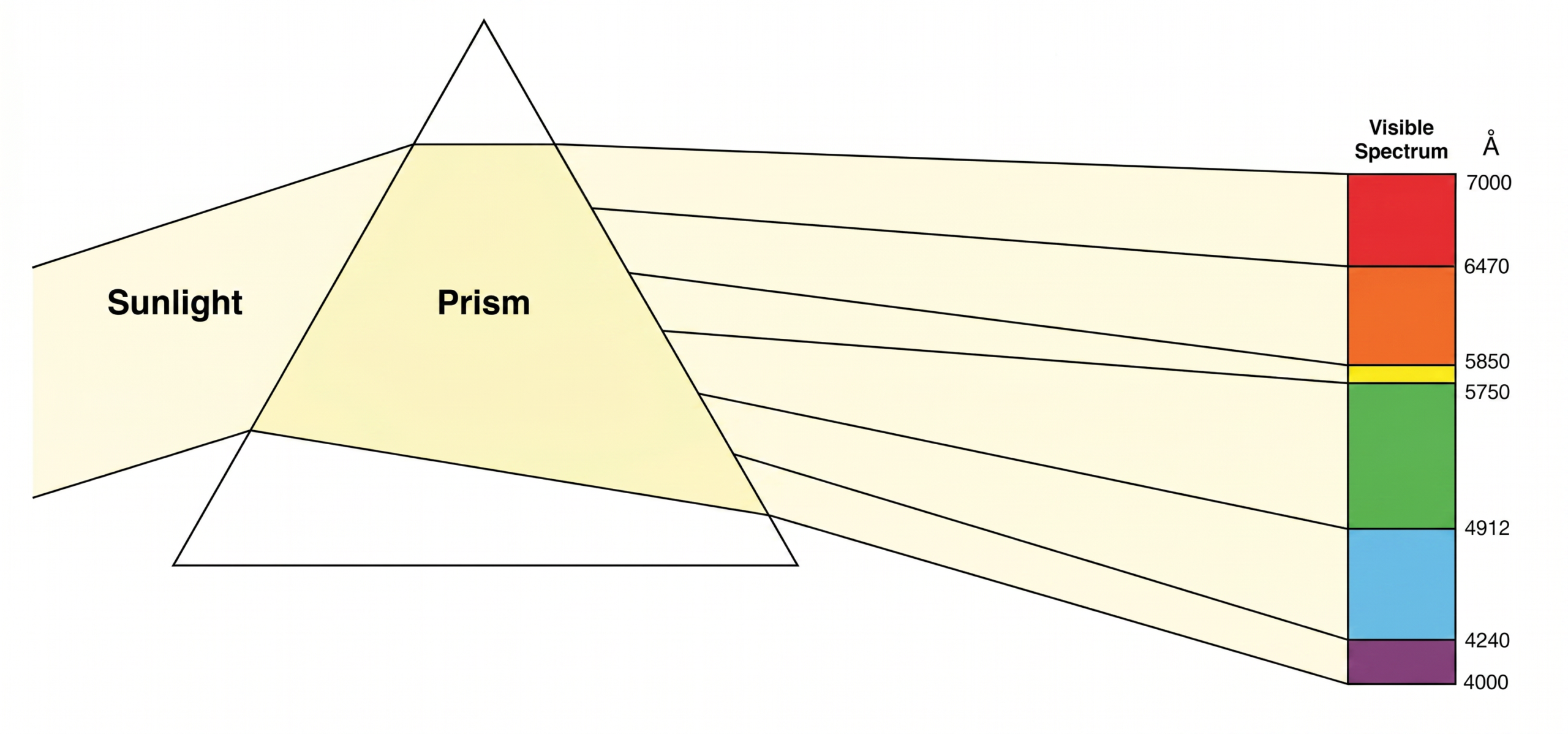
Light is a form of electromagnetic radiation characterized by waves or rays of various wavelengths. The different wavelengths of the rays emanating from any light source determine its color. The human eye can see wavelengths within only a fairly narrow range, called the chromatic visible spectrum. When sunlight passes through a prism (Fig. 1), the light rays are separated into the six distinct color components of the visible spectrum—violet, blue, green light, yellow, orange, and red. The wavelengths of these light rays go from 4000 Å (violet) up to 7000 Å (red), the human limits of color perception.
Objects reflect some of the wavelengths of light while absorbing others, which is the reason for all the different colors that we see. A red rose, for example, reflects the red light part of the spectrum and absorbs the short wavelengths, such as blue light. White objects reflect all wavelengths equally. In contrast, black objects absorb most of the light rays.
The optics of the human eye involve rays of light entering the eye through its anterior opening, the pupil, and are focused by the lens so they meet at a single point on the light-sensitive inner lining (retina), specifically the fovea, of the eyeball. The optic nerve connects the retina to the specific portion of the brain (visual cortex, via the lateral geniculate nucleus) where visual images of color and dimension are formed within the complex visual system, transmitting vital nerve impulses.
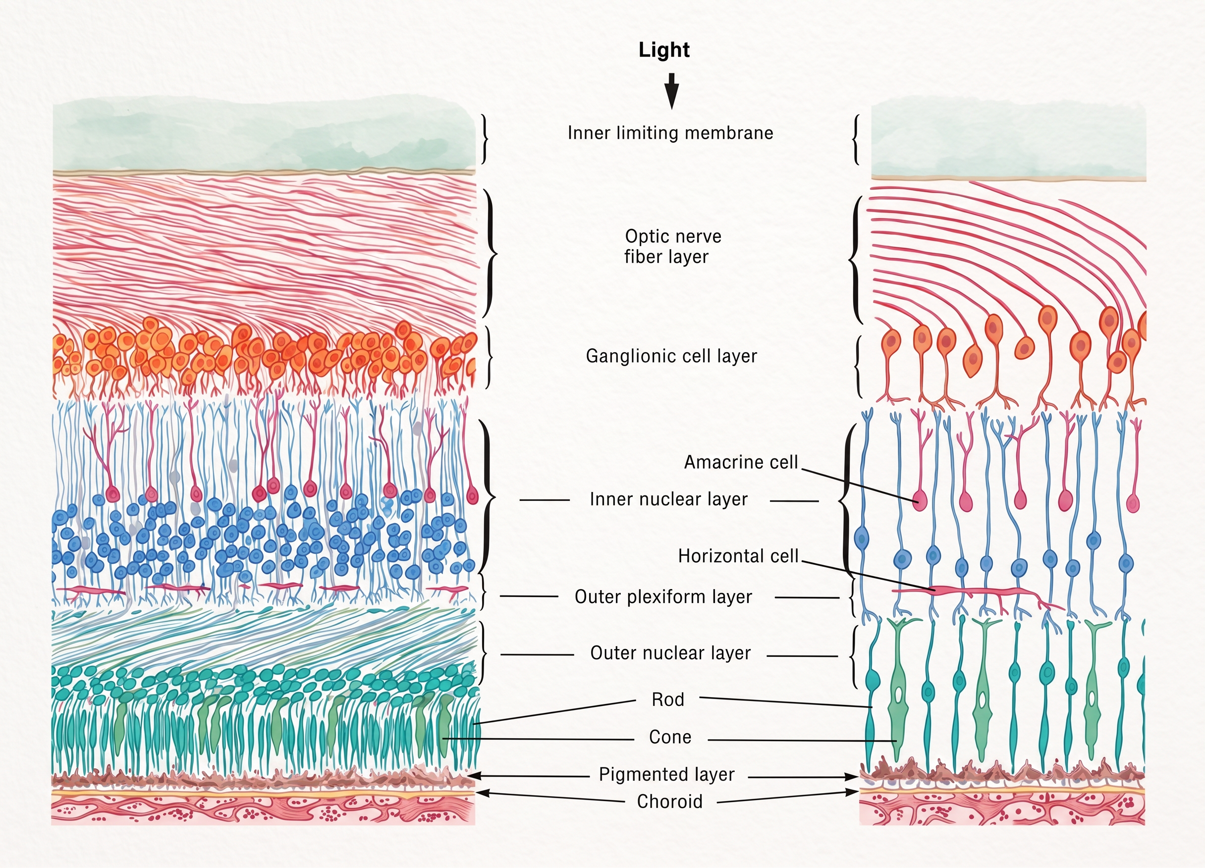
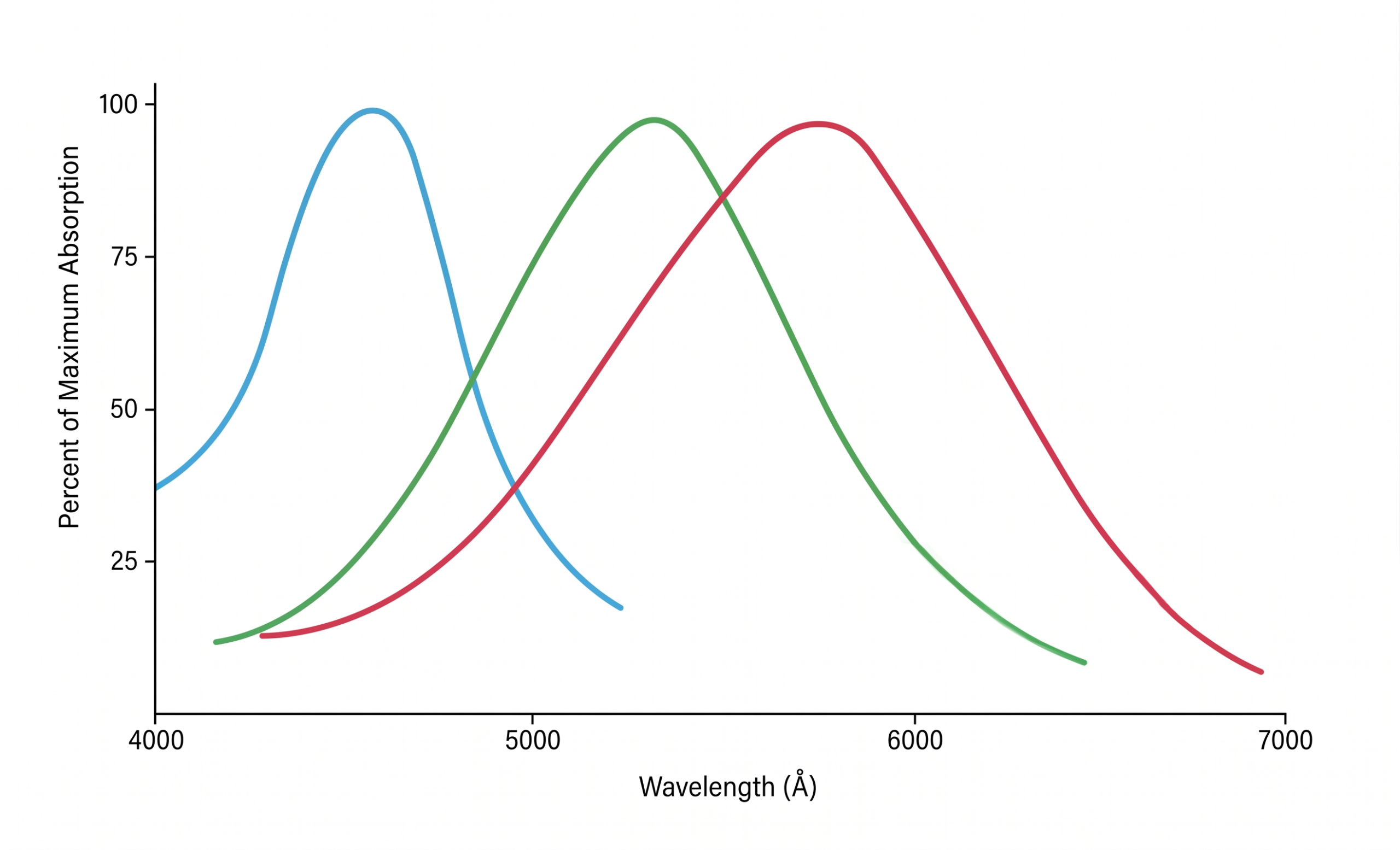
Within the retina are two types of nerve cells, or neurons, that are the receptors for light. Because of their distinct appearance, these photoreceptors are called rods and cones (Fig. 2). The rods are mainly responsible for black-and-white vision, especially in low light and varying light levels, and the cones for color vision. There are actually three types of cones (including L cones and M cones), each sensitive to a wavelength of light that corresponds to one of the primary colors (red, blue, and green). The responsiveness of each type of cone cell is decided by the specific light-sensitive pigment, or photopigments, that it contains, influencing its luminance perception. The sensitivities of the retinal cone cells are not exactly to the specific wavelengths of the primary colors, allowing for considerable overlap (Fig. 3). A person with normal color vision can differentiate hundreds of hues because of the stimulation of the various types of cones in specific proportions by different light stimuli, a phenomenon often linked to color constancy. Equal stimulation of the three types of cones is perceived as white light.
Color-vision deficiency, also known as color blindness, is the inability to distinguish certain shades of color. Persons with abnormal color perception may be classified into two general groups. Those who are totally color-deficient either lack active cone receptors entirely or have a defect in the visual cortex of the brain. The ability to distinguish the various wavelengths of light responsible for color vision is completely absent in these people, who are termed monochromats. The second group, called dichromats, characterized by dichromacy, have varying degrees of color-perception disability. They may be red-green or blue-yellow color-deficient, although the latter type is relatively rare. People with normal color vision are called trichromats because they exhibit trichromacy, being receptive to all three primary colors.
Red-green deficiency in color vision is divided into two categories according to the cone pigment abnormality involved. If the green cone pigment is deficient, the condition is the deutan variety. Affected individuals can distinguish only 5 to 25 color hues, all of which are perceived as shades of blue and yellow. An abnormality involving the red cone pigment is the protan type, which decreases the ability to distinguish the longer wavelengths of light (including long-wavelength red and orange).
Color-vision deficiency can be inherited or acquired through injury, disease, or certain types of poisoning. The inherited type affects both eyes equally in most cases and remains the same throughout life. Acquired color deficiency usually occurs in one eye more than the other and can become progressively worse, depending on its cause.
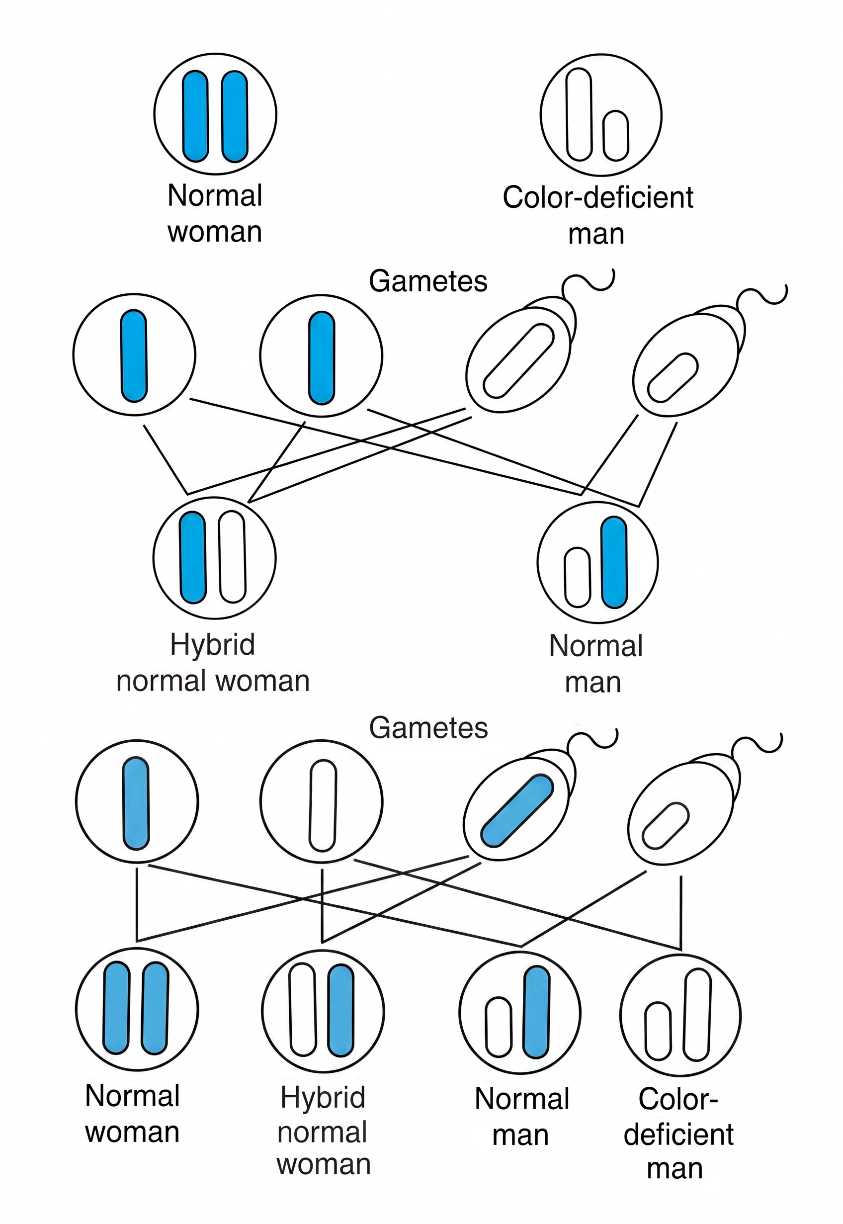
Hereditary color-vision deficiency is a sex-linked recessive trait which affects approximately eight percent of human males. The incidence in females is much lower (less than half of one percent). This discrepancy is caused by the way the genes for color discrimination are passed from parents to offspring, as shown in Figure 4. The gene for color-vision deficiency is carried on the X chromosome; males carry no color-vision genes on the Y chromosome. This gives the color-deficient male no normal gene to mask the expression of the abnormal gene carried on the X chromosome. He passes the recessive gene for color-vision abnormality to all of his daughters, who are then carriers of the deficiency but have normal color perception (unless the man’s mate is also a carrier of the recessive gene). According to the laws of probability, half of the daughters’ sons will be color deficient.
Although color-vision deficiency is usually not a significant handicap, there are important reasons in ophthalmology to test for this problem. A person who is unable to discriminate visual information readily, for example between red and green visible light, may be unable to follow traffic lights and shouldn’t drive a motor vehicle without special training. Abnormal color perception may be a limiting factor in certain career choices, like in the arts. Some color-deficient people need help selecting clothing. Color vision testing can be accomplished in the classroom quickly and easily while studying vision physiology.
One of the best tests of human color vision is the booklet Ishihara Standard Pseudoisochromatic Plates. This set of printed forms containing numerals can be recognized only by individuals with normal perception of certain colors. The subject examines all plates in the booklet and records the numeral that appears in each, effectively mapping their individual color space perception. Correct interpretations of the various colored plates are included with the testing materials. In addition to revealing color-vision deficiency, the Ishihara test separates the deutan and protan types of spectral sensitivities.
Another excellent way to test for color perception is the Holmgren’s wool test. A set of wool strands, each dyed a specific color, is mounted in a folder. The subject attempts to match each strand with an identical piece of yarn. Mismatching or hesitation in placing the strands correctly shows probable color-perception deficiency. This test is especially useful in mass screening, like in a classroom situation.
This article was originally published as “Human Color Vision” in Carolina Tips®, Vol. 43, No. 12 (print version, December 1980); it was revised April 2026.
Further Reading
Color blindness: holes in the rainbow. Current Health, 1980, 4(2), 24-25.
Hammond, Ronald E., Human Vision. Carolina Biological Supply Company, Burlington, 1980.
Martin. R., What it means to be color deficient, Innovations in Sight, May, 1979.
National Eye Institute. 2025. “Color Blindness.” https://www.nei.nih.gov/eye-health-information/eye-conditions-and-diseases/color-blindness.
Rushton, W. A. H., Visual pigments in man. Scientific American, 1962, 207, 120-132.
Wagh, Manasee. 2023. “How Colorblind Glasses Could Help You See More of the World.” Popular Mechanics, October 24. https://www.popularmechanics.com/science/health/a45442769/what-are-colorblind-glasses/.
Ronald E. Hammond, Ph.D.
From the Physiology Department,
Carolina Biological Supply Company, Burlington, North Carolina 27215
Get the latest news, free activities, teacher tips, product info, and more delivered to your inbox.