Mormoniella, a wasp slightly smaller than Drosophila, can be used for genetics studies with almost no preparation or equipment. It parasitizes fly pupae, which can be stored in a regular refrigerator for months. The wasps, with a ten‑day generation time at 30°C, can be reared in simple cotton‑stoppered vials, avoiding the need for culture media. Because males are haploid, offspring from unmated females show gametic ratios directly, making recessive mutations easy to detect without dominance masking.
The wasp will parasitize any fly pupae that are large enough. We have been rearing them on a large black and white striped fly of the genus Sarcophaga. Both the wasp and the fly are ideal for lab coursework in entomology and general biology. Their host-parasite relationship is fascinating, in addition to the uses that may be made of each species separately—unlike the microscopic interactions of protozoa—particularly in the study of beneficial insects like the braconid wasps such as Cotesia glomerata.
This genetics study covers these two insects, including these parasitoid wasps, which are now made available at all seasons of the year. They are both representatives of the Holometabola, insects with complete metamorphosis, in contrast to the Hemimetabola, such as grasshoppers, roaches and the sucking bugs, which have an immature or nymphal stage resembling the adult except for lack of wings.
The wasp and the fly hatch from the egg as legless larvae and retain this form until they reach full growth and pupate. The pupae are motionless externally, but within they undergo complete change finally eclosing as imagoes with all structures necessary for their new life. Moreover, their instincts are entirely changed.
The larva of Sarcophaga is without a true external head, acephalous, and is devoid of legs, apodous. Such larvae are known as maggots. At the pointed anterior end, there are two black mouth hooks used in feeding, and, just back of them on either side, the anterior spiracles that open into the tracheae or respiratory tubes inside the body. At the posterior end, inside a pocket, are the posterior spiracles, each with three split-like openings. Ventral to this recess are two fleshy projections between the anal opening.
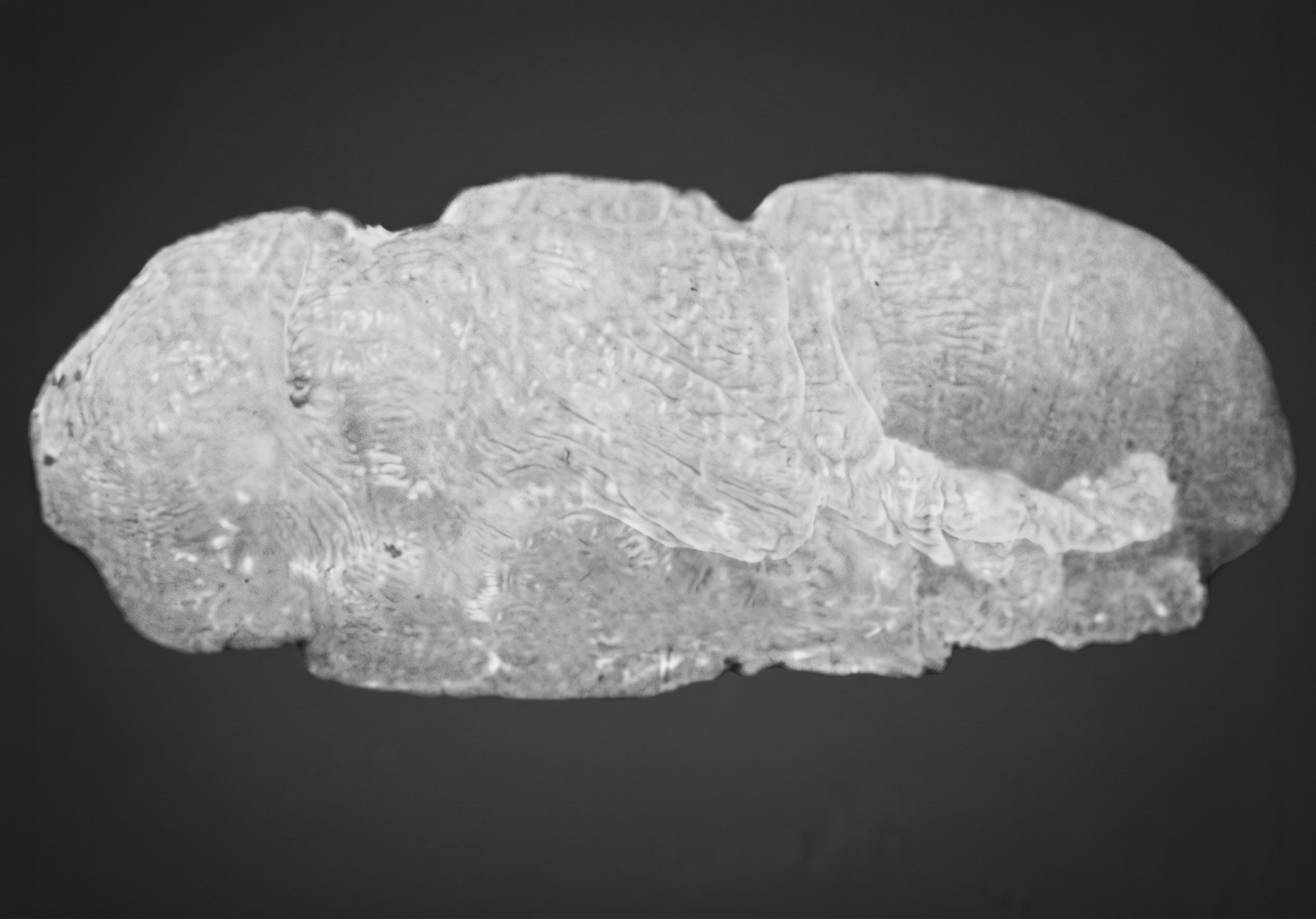
The larvae will develop to full size and leave their food—fish, meat, garbage—in eight days at a constant temperature of 26°C (79°F). They then bury themselves in the soil where pupation occurs. The integument of the larva (by darkening, hardening, and contracting to an oval shape) becomes the puparium or pupal covering. It typically takes 2 days from the time that this contraction takes place before the pupa is fully formed and separated from the puparium, leaving a free space between.
At the posterior end of the puparium is a pit containing the posterior spiracles or breathing pores of the larva. On either side of the anterior tip is a small projection called the anterior spiracle. The inside of the puparium shows the longitudinal glistening white larval tracheae, breathing tubes, and at the anterior end, the black mouth hooks of the larva. A puparium formed by a well-fed Sarcophaga maggot is about one-half inch in length.
If the puparium is held at its posterior end under a dissecting microscope, the anterior end may be carefully removed with a fine needle or sharp forceps. With a little practice, the entire pupa may be removed for study without rupturing its soft integument. Pupae may be stored for several weeks in an ordinary refrigerator and will retain their ability to develop when temperature is raised. The pupa resembles the adult fly in form except that the wings are enclosed in short ‘‘wing-pads.’’ It is at first creamy white but later takes on the color of the adult, or rather, the adult that forms inside the transparent pupal skin gradually takes on its definitive coloration. It takes about four or five days at 26°C from formation of the pupa until the eyes turn yellow, the large compound eyes at the sides and the small ocelli on top of the head. After another day, black bristles appear over the white body surface. Legs and the unexpanded wings become black and then the pigment spreads over the body. By removing puparia from the refrigerator on successive days until the first removed are ready for eclosion, it is possible to have pupae at the same time and to watch the process of eclosion.
The adult fly ecloses from the transparent pupal integument and emerges from the puparium. From yellowing of eyes to eclosion takes about five days at 26°C. When fully expanded, the fly appears much larger than the puparium where it emerged. This is because its tracheal sacs have been filled with air. The body of the fly is therefore light for its volume. It will float on water while the pupa will sink. However, the puparium containing the pupa will float because there is enough air space around the pupa to buoy it up.
Eclosion of the fly is an interesting process to watch. The anterior end of the puparium is split and forced off by expansion of a membranous “frontal” sac (ptilinum) that is thrust out from the head of the young fly. Alternate expansions and contractions of the ptilinum also enable the fly to force its way upward through the soil in which pupation has occurred. A freshly eclosed fly presents a rather spidery appearance running about with its long legs and small body. Shortly after eclosion, the ptilinum is withdrawn into the head, leaving only an inverted V-shaped mark, the frontal suture.
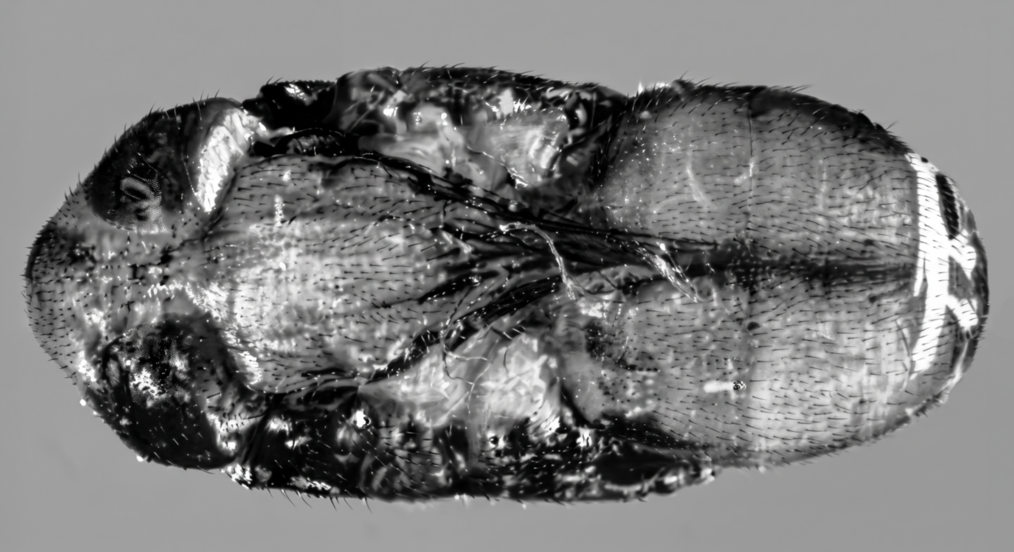
After resting a few minutes, the fly may increase in size and its wings may expand to full length. The entire process from eclosion to full expansion occupies about 15 minutes. The integument of the fly is now pale in color and soft in texture. The insect is therefore called teneral. Darkening and hardening takes place rapidly so that in less than one hour, flight is possible.
Mating takes place three to four days after eclosion. The eggs are fertilized and the embryos develop into maggots. Sarcophaga is larviparous rather than oviparous, meaning that the eggs hatch before they are laid, so that maggots rather than eggs are blown upon the food material.
Adult Fly Anatomy
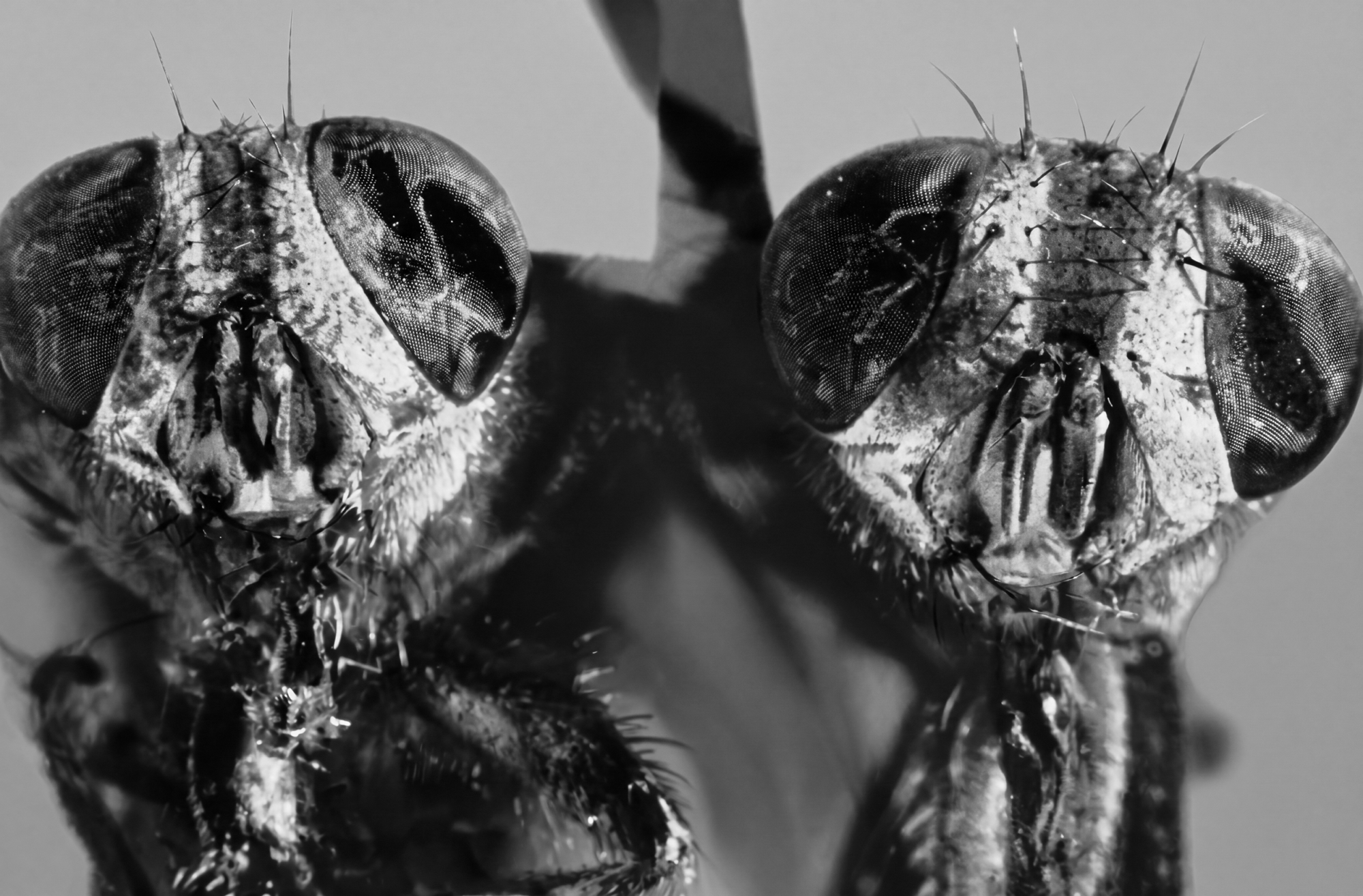
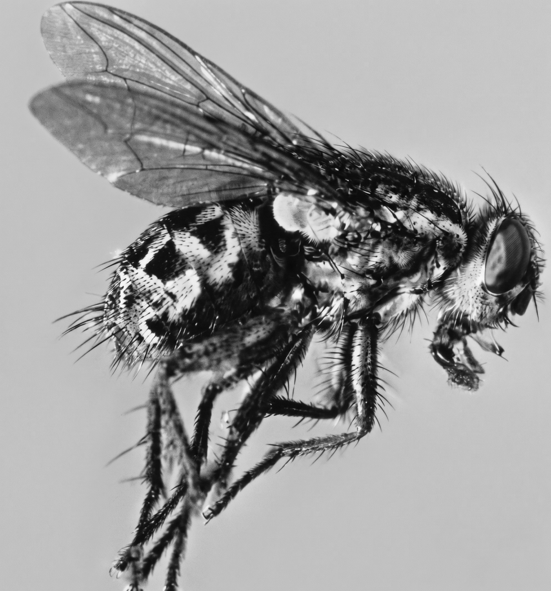
The body of the adult fly is divided into three definite regions—head, thorax, and abdomen. The head bears two large compound eyes, three small ocelli, an elongate jointed proboscis with two maxillary palpi pointing forward, and two antennae consisting of three segments—two small and one long terminal. A bristle-like structure, the arista, somewhat plumose, projects from near the base of this terminal segment. This has evolved phylogenetically from the many-segmented antennae of more primitive flies such as gnats and mosquitos. If the head of a fly is removed and placed flat upon a slide, it is convenient to study these structures and also observe the inverted V-shaped ptilinal suture.
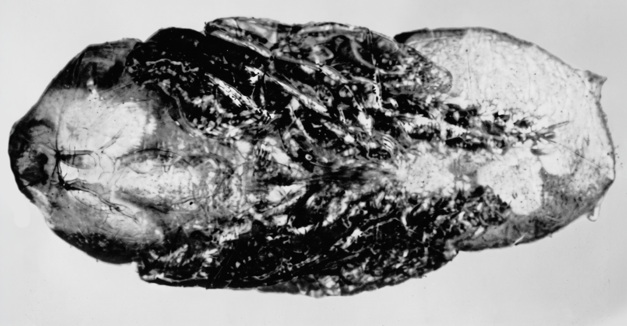
The thorax consists of three body segments corresponding to the three pairs of legs—prothorax, mesothorax and metathorax. The metathoracic pair of wings found in other insects is replaced in flies by elongate balancing organs, halteres, protruding posterior to the wings.
In the Sarcophaga group of flies, there are two flat scale-like structures posterior to each wing—the squamae or calypterae, which may cover the halteres.
Each leg is attached to the body by a region called the coxa. The small segment at the base of the leg is the trochanter which is followed by the elongate femur and tibia and the five-segmented tarsus, the terminal segment bearing a pair of claws with a cushion or pulvillus between them. Dorsally the prothorax and metathorax are barely visible, being much reduced while the mesothorax is expanded. This dorsal part, called the mesonotum, is differentiated into three conspicuous regions separated by two transverse sutures. Under the mesonotum are situated tracheal air sacs.
What are called direct wing muscles are small and used for folding the wings back when not in use, also for steering during flight. The bulk of the work of flight is carried on by relatively large muscles, not attached to the wings, and known as indirect wing muscles. These may be easily seen if a preserved fly is cut transversely with a. razor blade just in front of the wings. Centrally, toward the dorsal side, are two bundles of muscles that run longitudinally (seen in cross section). These depress the wings by producing a longitudinal arching of the mesonotum. On each side, there are large bundles of muscle fibers that run vertically (seen in side view). These tergo-sternal muscles elevate the wings by drawing down the arched back.
It is interesting to observe an adult fly feeding. If a small drop of sweetened water is placed into a vial containing a hungry fly, the tongue with its two long maxillary palpi will be extended and the fluid drawn up. Fly food must be dissolved or suspended in water. Flies have a special sucking stomach, known as a crop, in the abdomen for storing food until it is digested. This corresponds to the honeybag of moths and butterflies. By expansion of this crop liquid food passes through the tongue and the long esophagus in the thorax. A rasping organ at the tip of the tongue enables the feeding fly to scrape off particles of solid food which are then mixed with fluid. While observing this, one may also note the five-segmented tarsi with their claws and pulvilli.
Sex is determined by the width of the head between the eyes and by the genitalia. In many flies the compound eyes of the male are very much larger than those of the female so that they meet dorsally. The males of such species are said to be holoptic, while the females are dichoptic. In Sarcophaga both sexes are dichoptic but the space between the eyes of the female is wider and within this space there is a bristle on either side of the antenna, the orbital bristle, not present in the male. The female also has an extra bristle on top of the head, the outer vertical.
The genitalia of the male are very elaborate. Species identification within the Sarcophagidae is based largely on a study of these structures and there has been no corresponding evolution within the females. In fact, species identification is often very difficult or impossible if females only are available.
Other sex differences are present but are inconspicuous or hard to define. Differences in body shape and curvature of certain parts of the legs may be noted.
As with other species of flies, the size of the adult is determined largely by the opportunity afforded the larvae for feeding. All growth occurs during the maggot stage and there is often severe competition for food. Small flies are not young flies. Flies in general are adapted to mature in spite of greatly reduced rations.
The insect order Hymenoptera includes bees, wasps, and ants, social insects of popular interest, but the vast majority of species are not social—unlike parasitic flukes or other helminths, these include saw flies and gall wasps feeding on plant material, ichneumon flies attacking caterpillars, larvae and pupae of many insects and a vast host of egg parasites belonging to different superfamilies. The largest superfamily of the order is the Chalcidoidea or chalcis flies, including Encarsia formosa, for the most part minute egg parasites associated with various parasitic infections, hyperparasitoids, or specialists attacking aphids. Mormoniella, although a small insect fluctuating about two millimeters in length—significantly larger than a single-celled Plasmodium—is one of the larger of the chalcis flies. The species is worldwide in distribution, including regions affected by neglected tropical diseases and other forms of parasitic disease, and may be found wherever the larger species of flies are forming puparia.
The female wasp stings the fly pupa through the puparium. The pupal juice, oozing out, forms a tube connecting the wound with the opening made in the puparium and it is through this tube that the wasp feeds. If a portion of the puparium near the puncture is removed, a black spot will be seen to have formed in the body of the pupa and frequently a trace of the tube may be found near the center of this spot. A well-fed female develops her eggs, oval and creamy-white in color, and deposits them through her sting, which serves also as an ovipositor, in the space between the pupa and the puparium.
Up to 20 eggs may be laid about a single puncture and the same female or other females may parasitize the same host pupa, feeding and ovipositing in different regions. The eggs hatch into legless larvae that, in contrast to the maggots of flies or the simple structure of nematodes and other roundworms, possess a small head and have spiracles arranged in a row on each side of the body. These larvae, inside the puparium, lie and feed upon the outside of the pupa as ectoparasites.
When full-grown, the larvae transform into pupae still within the host puparium. These pupae at first are white but later darken becoming almost black in color. At this stage, sex may be readily identified. The females, averaging somewhat larger than the males and being somewhat more convex dorsally, show a median white streak ventrally on the abdomen, the region of the differentiating sting or ovipositor. If a well-parasitized puparium be broken open, there may be removed from it many wasp pupae and the shriveled remnant of the fly pupa.
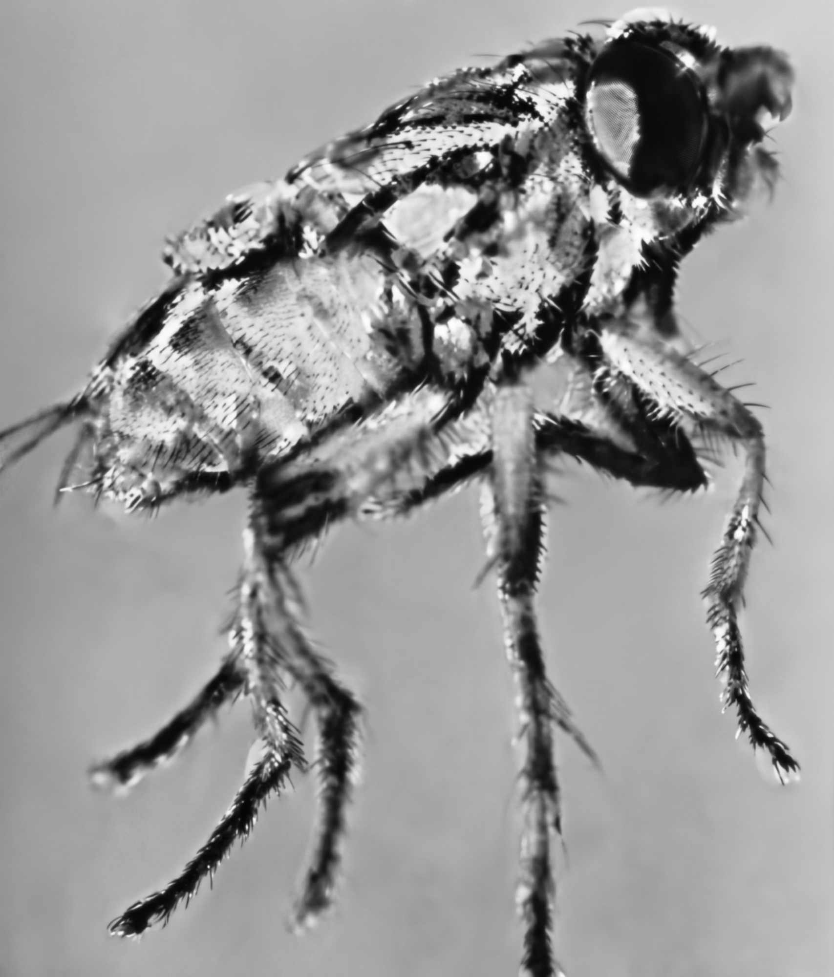
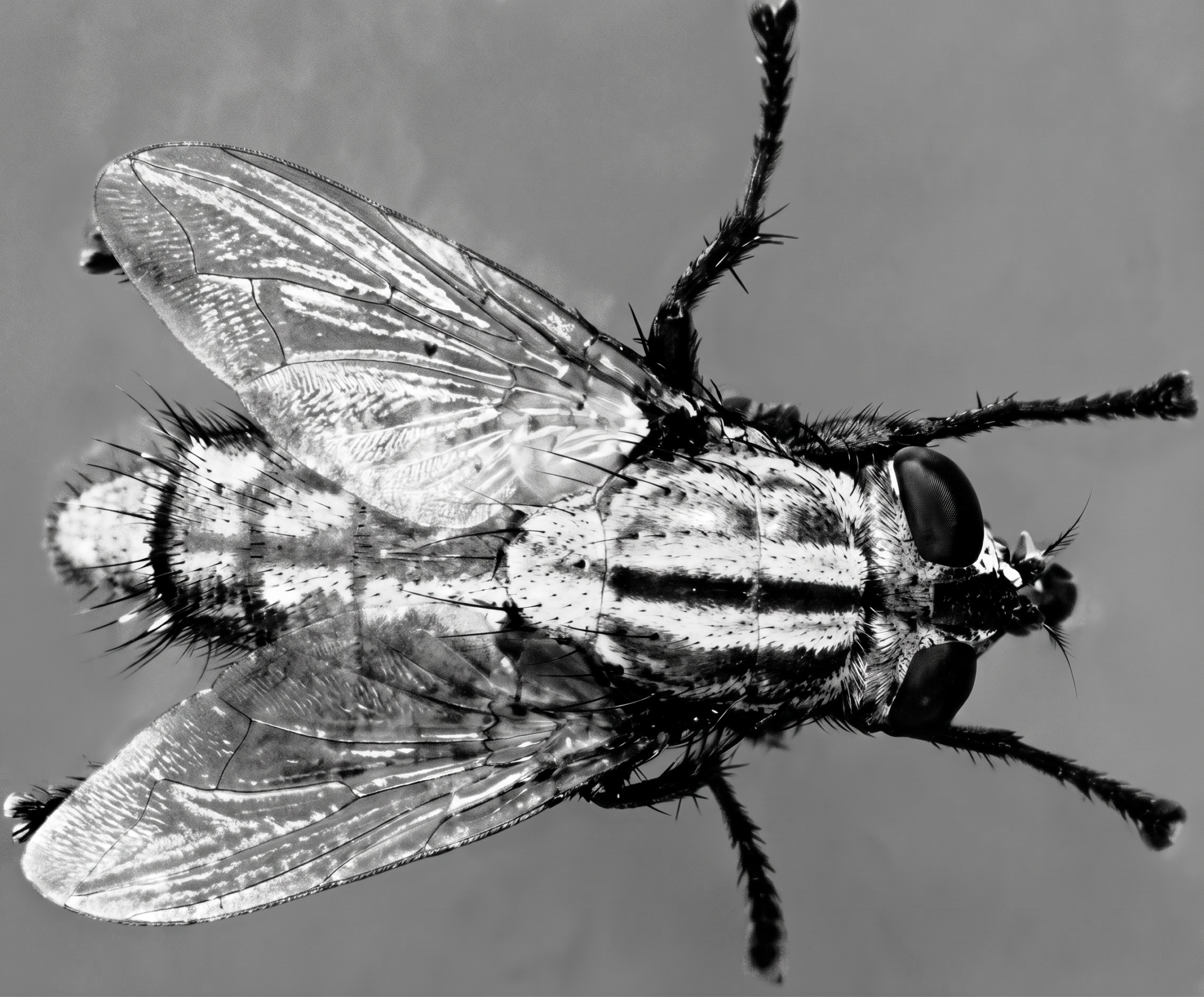
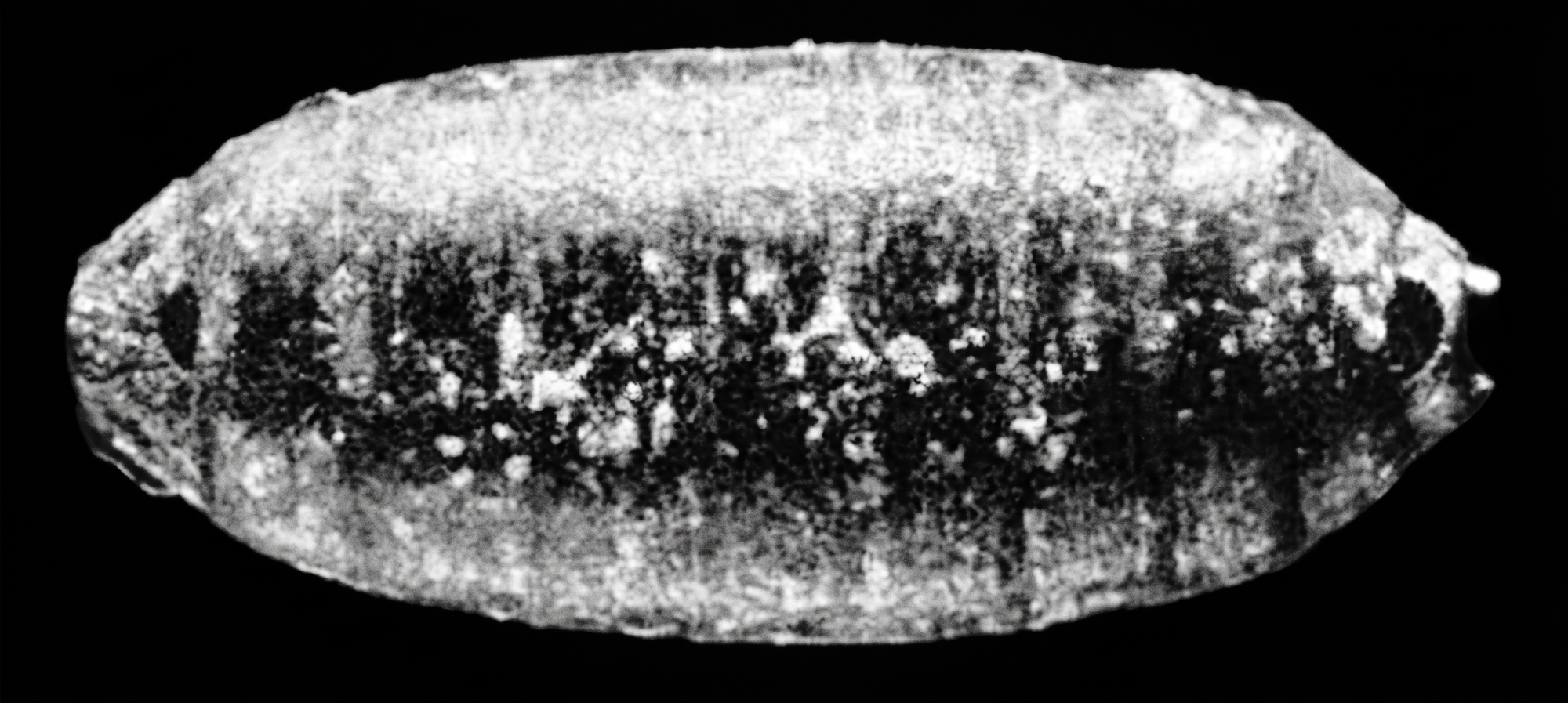
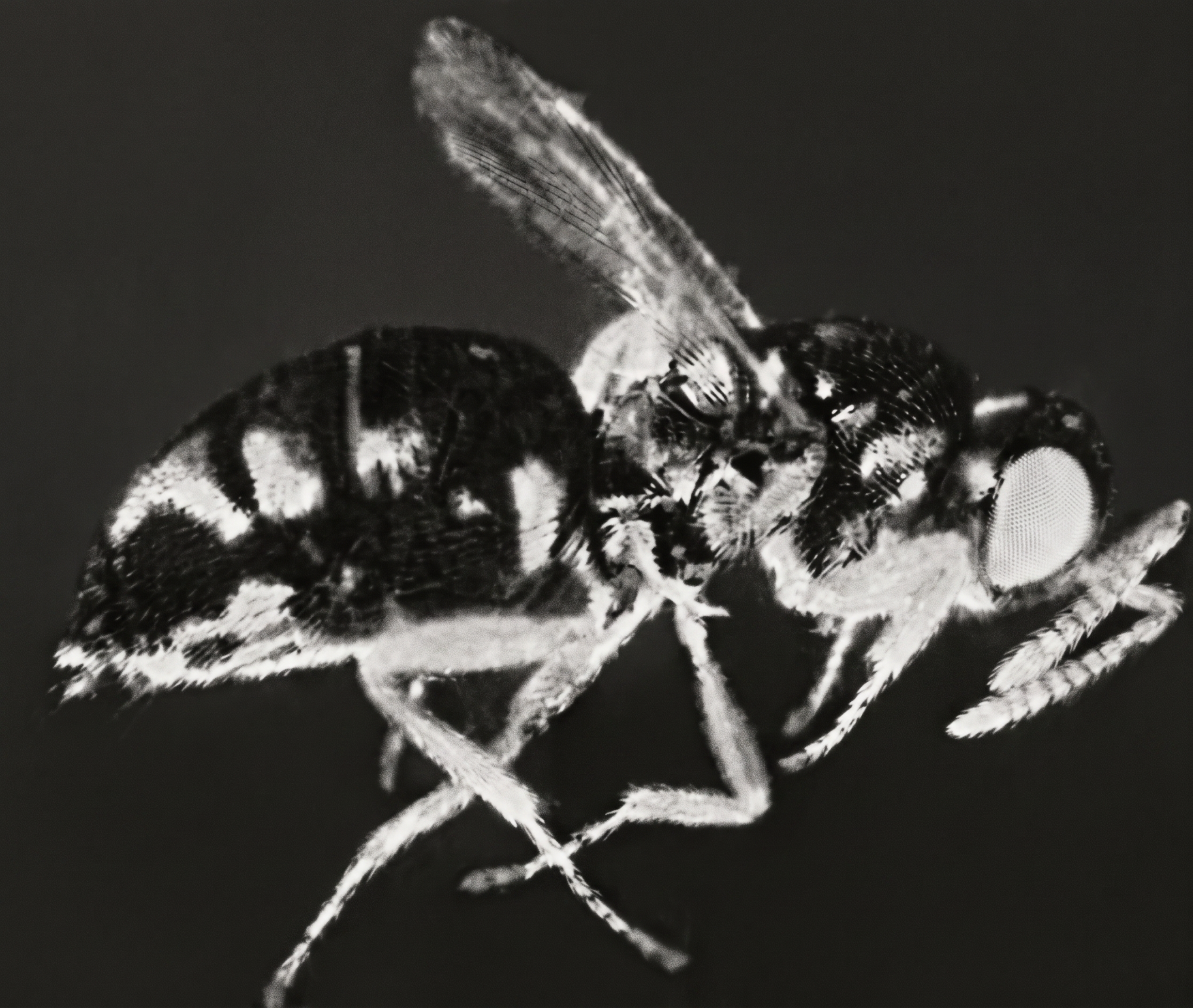
Mormoniella, like its host Sarcophaga, is adapted for overcrowding. By setting several females with a few pupae it is possible to obtain over 200 individuals from a single large puparium. These will be exceedingly small. The optimum number for full growth, utilizing all the food material provided by the body of the host pupae, is from 50 to 75.
The active imagoes eclose, leaving the thin pupal exuviae, much like other Hymenoptera emerging from cocoons. The fly puparium may be packed with wasps if it is well parasitized and almost all of them may be adults if the eggs were laid at approximately the same time. These wasps may include both sexes if the mothers were mated, but from unmated females there are obtained only males. One or more holes may be gnawed in the puparium through which the brood emerges. If the brood is bisexual, it is usually a female that gnaws the hole. Occasionally a puparium will be found filled with dead and dried adults. These are usually all males, none of which apparently had initiative enough to gnaw the hole. Had this puparium been opened before the wasps died, they would have been found to be active and healthy.
Sarcophaga pupae can be refrigerated for long periods of time and will still serve as adequate food material for rearing Mormoniella. Pupae refrigerated over a year ago are still being used as hosts. They produce an abundance of healthy wasps, almost every puparium exposed being filled with offspring after incubation. When these pupae are incubated without being infected, they soon turn brown, die, and dry, with no flies developing from them.
A puparium from which adult wasps have emerged may contain in addition to the exuviae of the wasps and the remnant of the fly, one or more diapause wasp larvae. Diapause is a condition of dormancy which larvae may assume if subjected to cool temperature. Structurally a diapause larva remains immature, but growth continues and much fat is deposited until the body is frequently larger than that of a larva about to pupate. Larvae may remain in diapause for many months. Recently several groups of larvae that had entered diapause two years previously were taken out of the refrigerator and shortly pupated and matured into normal fertile adults. Diapause may be broken by keeping the larvae in a refrigerator for three months or more. If they are then placed in an incubator they will pupate. Diapause larvae will live as such for several weeks in an incubator failing to pupate unless refrigerated. Diapause has the effect of spreading eclosions of the progeny of single female over a considerable period of time. This must prove of adaptive value giving a much greater chance that the line may be perpetuated if food is available at rare intervals only.
Sex of adult Mormoniella is determined by the brighter body color of the male which has pale antennae. Females have black antennae, except for the basal segments. Wings of females reach approximately to the tip of the abdomen, while males have very short wings and are unable to fly. The females do not often fly; instead, they prefer to run about very actively and jump for considerable distances with their wings.
Mating normally takes place as soon as the wasps have emerged. In a culture vial, the males may be seen running about in frenzied search of females. This may continue for several hours. If the wasps are quieted with ether vapor for examination, the mating activity will usually be resumed immediately after recovery. However, two- or three-day-old mates will sometimes be altogether indifferent so that it is impossible to get matings. Females also mate readily shortly after emergence, but it has been possible, in a small percentage of cases, to cross a virgin female to her own son. This was accomplished by placing the female in a refrigerator while her brood was kept in an incubator. When males appeared, they were set with the virgin mother which then produced daughters.
Mormoniella is unusual because males are haploid—they develop from unfertilized eggs and have only one set of genes. This makes their genetics much simpler than in most organisms.
In typical diploid species, we often talk about the 3:1 Mendelian ratio (three dominant to one recessive in the F₂ generation). But this ratio is actually the result of combining egg and sperm, each carrying a 1:1 gametic ratio, plus the effects of dominance. It’s like tossing two coins at once and counting any toss with at least one “heads” as dominant.
Because Mormoniella males come from unfertilized eggs, we don’t see the 3:1 ratio. Instead, we see the true fundamental ratio in genetics: 1:1, directly in the male offspring.
The fundamental one-to-one Mendelian ratio may be readily demonstrated with Mormoniella by crossing two stocks differing by a single gene and breeding the offspring, all males, produced by unmated F₁ females. The “F1” males will really be of the pure maternal stock, coming from unfertilized eggs. They therefore have no fathers and are half-brothers to their sisters. Gametes produced by any one male are all alike; he breeds like a homozygote however heterozygous was his mother.
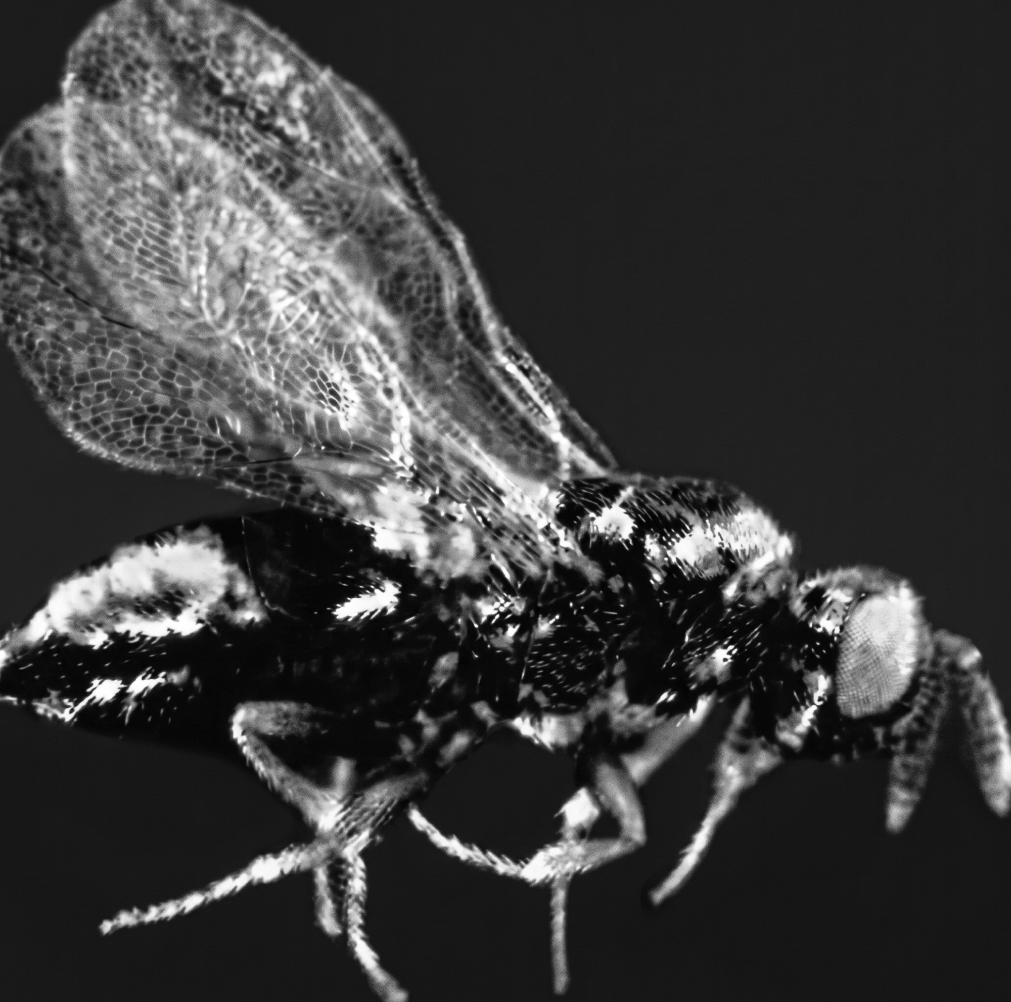
Many gene differences have been found in Mormoniella, originating in mutations both spontaneous and induced by radiation. In this species, most of the mutations are in eye color, several in body color, and a few in structural traits.
Wild‑type Mormoniella eyes are dark brown. All eye‑color mutations are recessive to this. Many mutant colors exist, mostly different shades of red—ranging from dark red (dahlia) to bright scarlet. Some mutations dilute pigment, producing lighter colors like tomato, peach, and finally oyster, which is almost completely colorless.
If a female is unmated and carries one mutant eye‑color gene, she produces sons in a 1:1 ratio: half wild‑type, half mutant.
Examples:
+/st → wild‑type and scarlet sons
+/oy → wild‑type and oyster sons
+/da → wild‑type and dahlia sons
+/vm → wild‑type and vermilion sons
If a female carries two different mutant alleles, her sons show both mutant types in equal numbers (e.g., st/oy → scarlet and oyster sons). This shows that all these eye‑color genes belong to one allelic series, called the R series.
Some eye‑color genes are not part of the R series. They produce similar colors but segregate independently and can interact with R‑series genes.
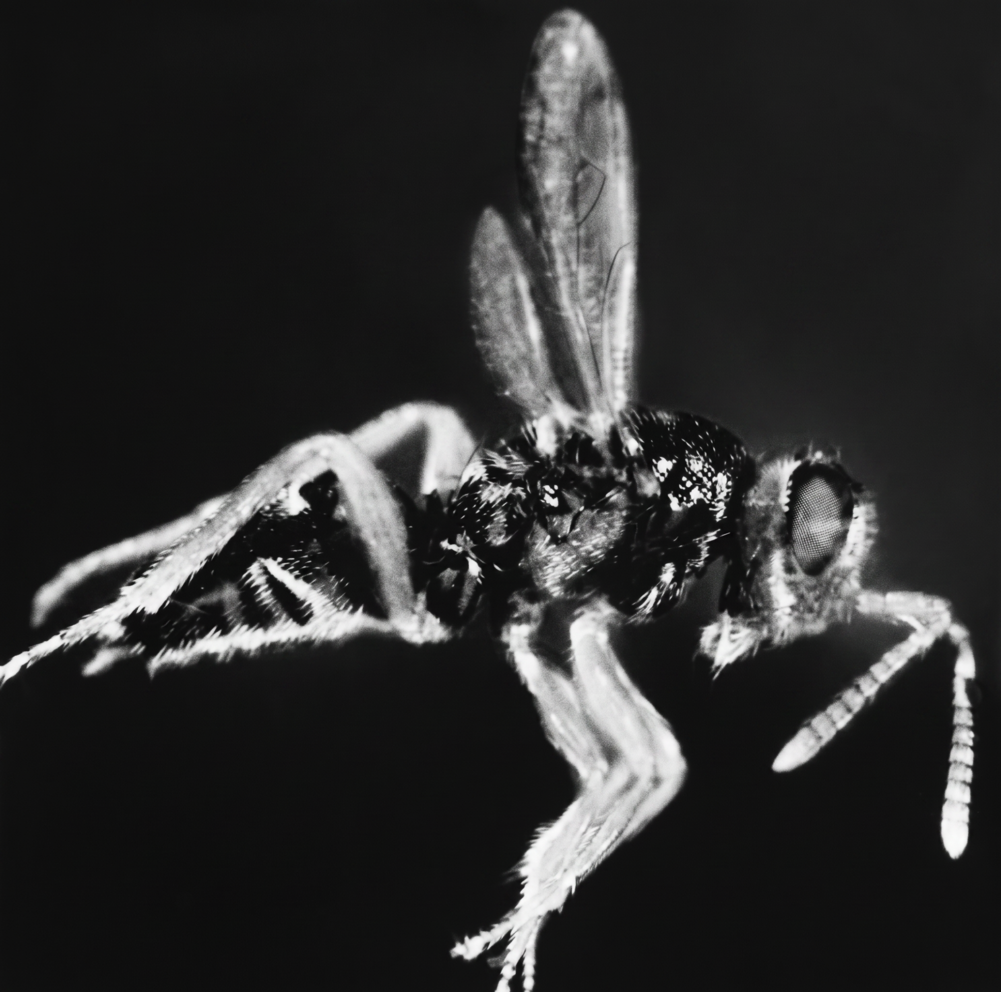
Wild‑type body color is bronze‑green. A mutation called purple is linked to the R locus.
Purple × red → F₁ females produce four male types: wild‑type, purple, red, purple‑red.
Purple × scarlet → linkage appears: mostly purple and scarlet sons, with a few recombinants.
The glass mutation reduces eye size.
Glass × red → simple Mendelian 1:1:1:1 male ratio.
Glass × scarlet → strong linkage (about 1% recombination).
Two mutations—black (bk) and ebony (eb)—produce black eyes but behave differently genetically. They are independent of each other and of the R series, but show epistasis (one gene masking another).
Examples:
dahlia + black → dark purple
scarlet + black → white
dahlia + ebony → near‑black
scarlet + ebony → scarlet (scarlet masks ebony)
vermilion + black → lavender
peach + black → oyster
peach + ebony → oyster
vermilion + ebony → vermilion
Black × ebony → F₂ males show a 1 wild‑type : 3 black ratio. Orange is used as a marker because orange‑black and orange‑ebony look oyster‑white.
Some R‑series alleles are lethal or female‑sterile.
Example:
scarlet lethal (stl) must be kept with oyster female‑sterile (oyfs).
stl/oyfs females produce only oyster males because stl eggs die.
oyfs/oyfs females are sterile.
Male wasps are haploid, so dominance doesn’t apply—they have only one allele. Females are diploid, so dominance, recessiveness, blending, and co‑dominance can appear. Some compound heterozygotes look wild‑type but produce only mutant sons.
Some alleles look identical but behave differently in combinations.
Example:
Oyster‑1 complements scarlet‑1, partially complements scarlet‑2, and is recessive to scarlet‑3.
Oyster‑2 is recessive to all scarlets.
Mormoniella shows clear examples of Mendelian inheritance, linkage, epistasis, lethals, and gene interaction. Because the wasps are easy to rear and males are haploid, they are excellent for genetics studies.
1. Aldrich, J. M., 1916. Sarcophaga and Allies in North America. The Thomas Say Foundation.
2. Curran. C. H., 1934. The Families and Genera of North American Diptera. The Ballou Press, New York, New York.
3. Hall, David G, 1948. The Blowflies of North America. The Thomas Say Foundation, Vol. 4. Monumental Printing Co., Baltimore, Maryland.
4. Lowne, B. Thompson, 1890-95. The Anatomy, Physiology, Morphology and Development of the Blow Fly. Vol. I, pp. 1-350, Vol. II, pp. 351-778.
5. Roback, Selwyn S., 1954. The Evolution and Taxonomy of the Sarcophaginae. Illinois Biological Monographs. Vol. 23, Nos. 3-4.
This article was originally published in Carolina Tips®, April 1955; it was revised July 2026.
Get the latest news, free activities, teacher tips, product info, and more delivered to your inbox.