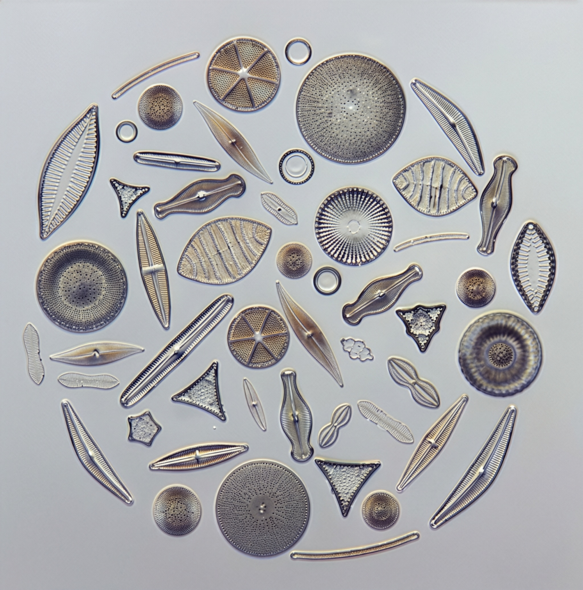
Diatoms are a diverse group of eukaryotic microscopic algae, classified as Bacillariophyceae and often referred to as single-celled algae, that play an essential role in aquatic ecosystems and the global carbon cycle. It is estimated that there are between 5,000 and 10,000 different diatom species found throughout the world in moist soil, oceans, freshwater rivers, lakes, and oceans. Diatoms are among the most important primary algal producers and protists on Earth, alongside groups like dinoflagellates, responsible for an estimated 20–25% of global photosynthesis. The cell walls of diatoms show an intricate beauty that is unsurpassed.
Diatoms have been a part of the world’s ecology since Mesozoic times. They have occurred in enormous numbers, leaving vast deposits of siliceous cell walls that are used today as a part of several hundred industrial products (e.g., chalk, talc, abrasives, filters). They support the base of food chains and sequester carbon dioxide, helping to stabilize climate change. This unique and beautiful species has also spawned an art genre, illustrating again the complexity, beauty, and scope of the natural world.
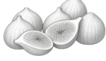
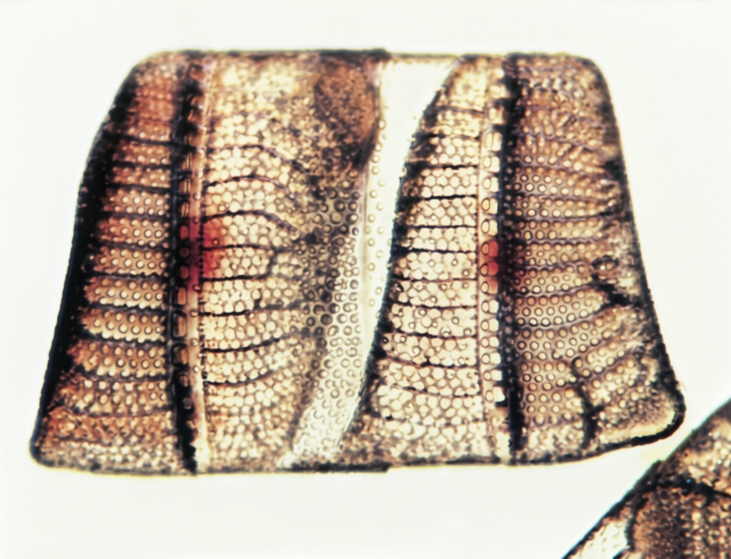
Diatoms live in “glass houses.” They have beautiful, ornamented, silicified cell walls. This is evident if the protoplast is removed by acid or heat (Fig. I).
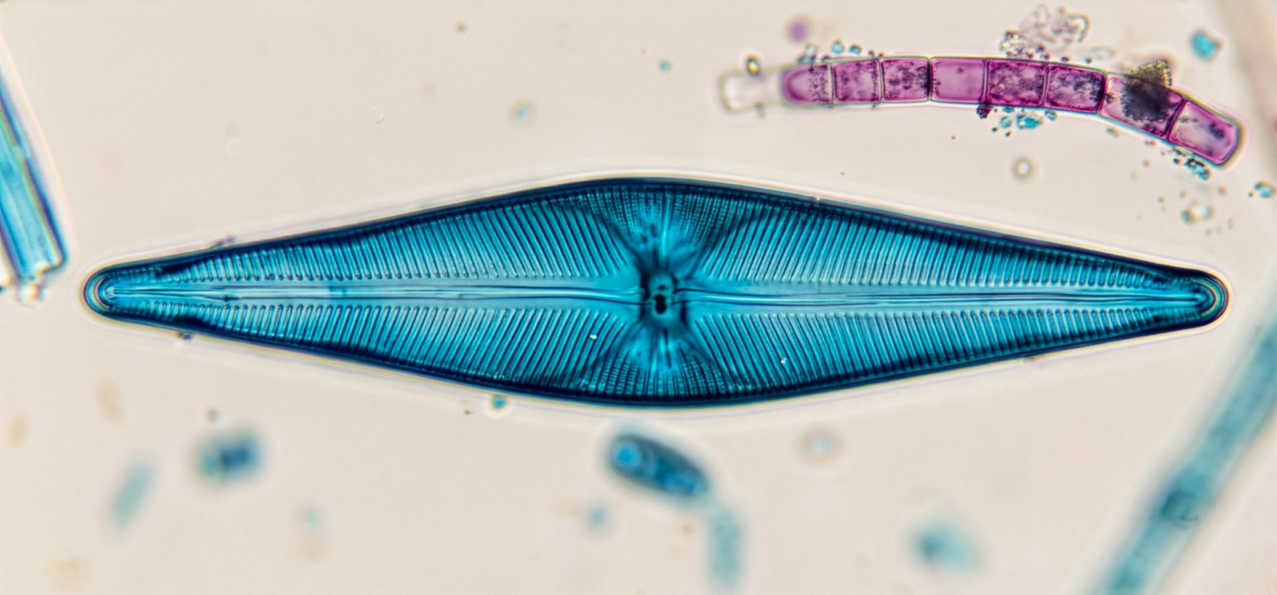
In most diatoms, the diatom cell wall, called a frustule, is made up of pectin impregnated with hydrated silica (hydrated silicon dioxide). Electron microscopy has shown that the fine markings of the diatom walls are actually pores which give the protoplasm access to the external environment. The walls consist of two interlocking halves, (valves) much like a petri dish or a gelatin capsule. These halves—called the epitheca and hypotheca—are often intricately patterned with pores and ridges that vary by species. Inside the frustule, diatoms contain chloroplasts with chlorophyll a and accessory pigments such as fucoxanthin, which give many diatoms a golden-brown color. The outer valve, the epitheca, fits over the inner valve, the hypotheca (Fig. 2). The two valves may be attached to a girdle band instead of overlapping. The view from above or below is called valve view (Fig. 3). The view from the side is the girdle view. Their unique structure not only provides protection but also aids in buoyancy and light absorption.
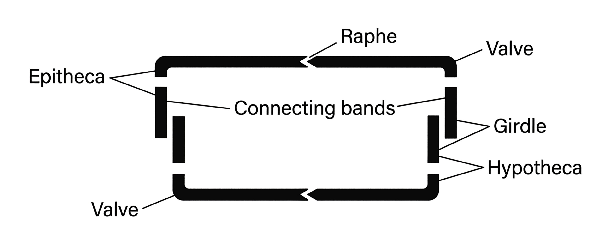
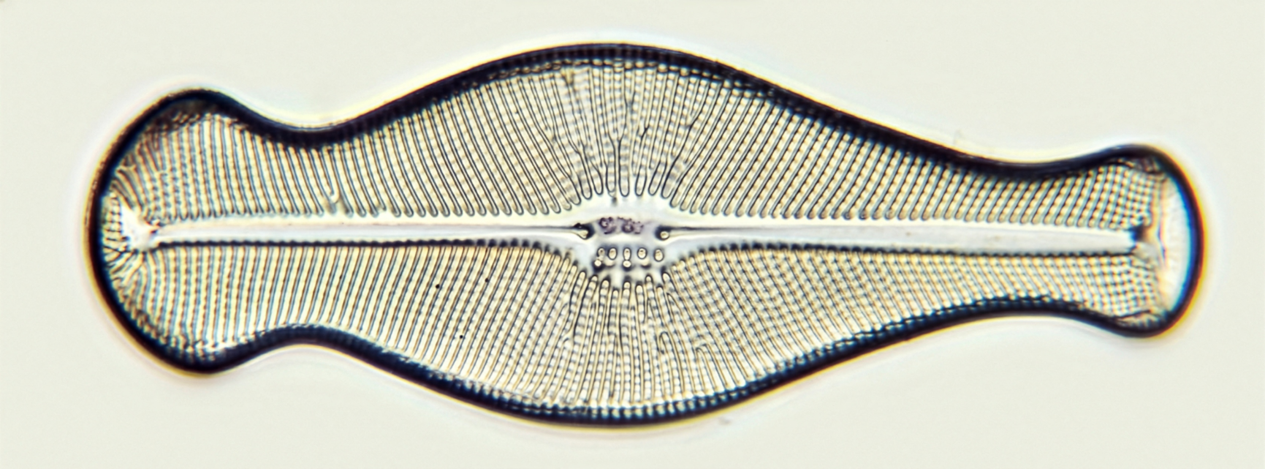
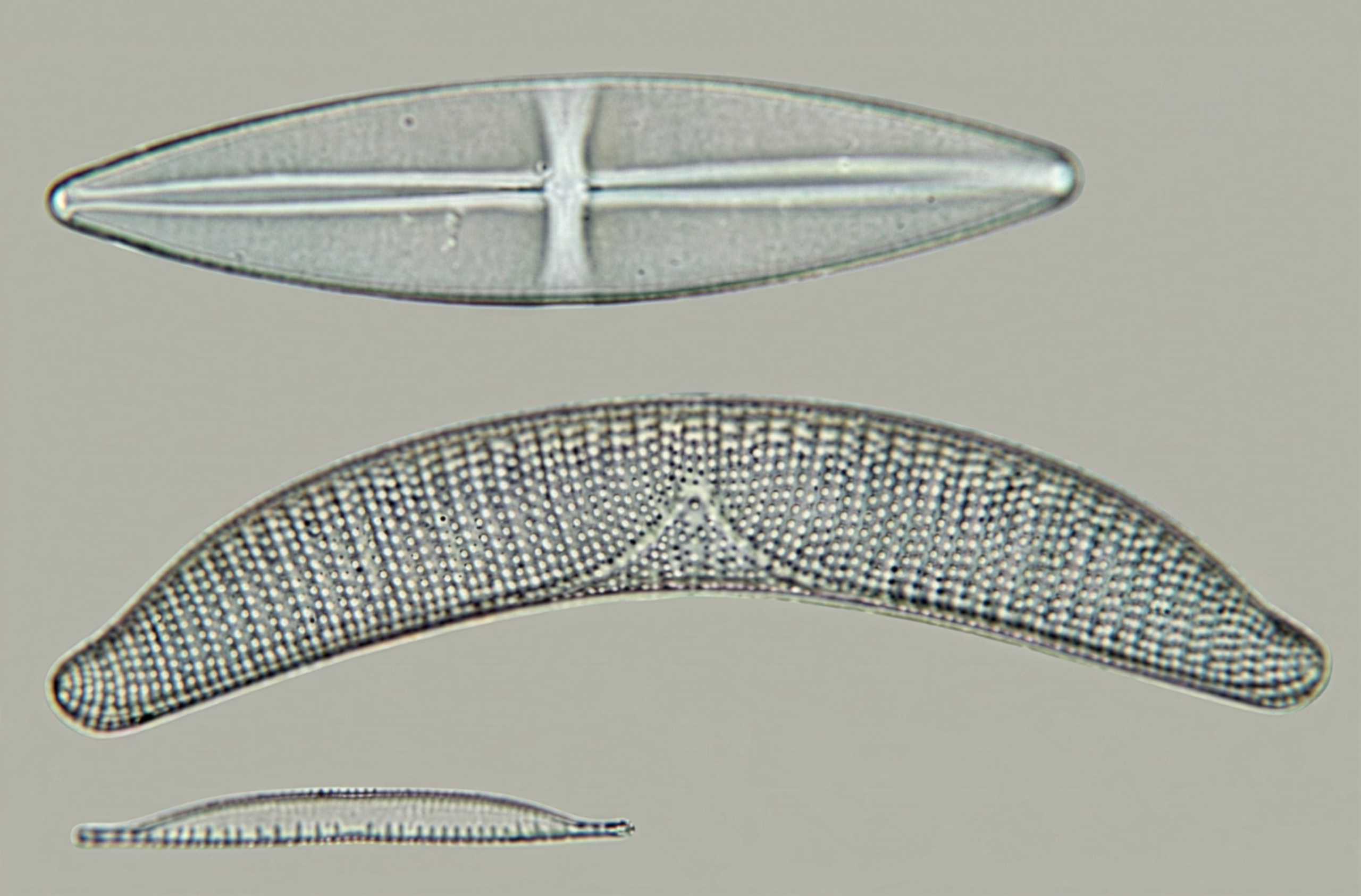
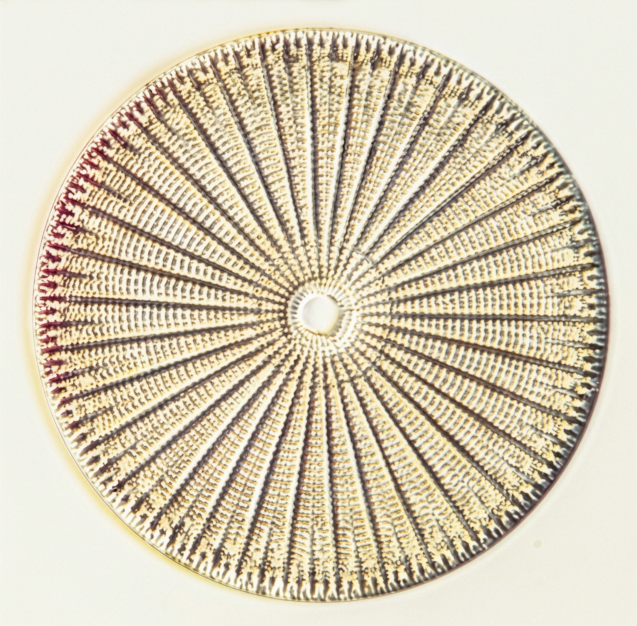
The valves are either pennate or centric. Pennate diatoms have bilateral symmetry in valve view. The general outline may be boat-shaped or rod-shaped (Fig. 4). In the center of the valve of most pennate diatoms is either an unsilicified groove, the raphe, or a hyaline median line, the pseudoraphe (Fig. 3). The raphe seems to be associated with the movement of many pennate diatoms. Centric diatoms have radial symmetry in valve view and lack raphes and pseudoraphes (Fig. 5). The outline of centric diatoms is usually circular, oval, or elliptical.
The protoplast of a diatom has a nucleus, a central vacuole, and one to several brownish plastids which contain chlorophylls a and c as well as 3-carotene and xanthophylls. Food is stored as oil or leucosin in vesicles in the cell. (Fig. 6).
Diatoms are found in both terrestrial and surface water environments, including freshwater and marine habitats, often coexisting with other microorganisms like cyanobacteria and various microbes depending on the environmental conditions. Marine diatoms and freshwater diatoms, which adapt to varying levels of salinity and even extreme conditions within sea ice, can live suspended in the water column (planktonic) or attached to surfaces such as rocks, plants, and sediments (benthic). They may be solitary or colonial. Some living diatoms secrete a mucilaginous capsule. The mucilage may hold groups of diatoms in patterns such as ribbons, long-chains, zig-zag ribbons, branched chains, and various other configurations.
Diatoms are important because they, along with green algae, other phytoplankton, zooplankton, and plankton, form the base of the food web and food chain for animals in both freshwater and marine environments. Additionally, because diatoms are highly sensitive to changes in water chemistry, temperature, and nutrient concentrations, they are often used as biological indicators to assess water quality and the impacts of climate change.
Asexual reproduction takes place by nuclear and cytoplasmic division (Fig. 7). The daughter cells each have only one wall and half the volume of the parent cell. As a daughter cell enlarges, a new wall develops, with the parental wall forming the epitheca. Thus, one daughter cell will be smaller than the parent cell. Continued asexual reproduction of this type results in the reduction in the average size of the individual diatom. Normal cell size is restored by discarding the old frustule and forming a new frustule over an enlarged protoplast, sometimes involving the formation of resting spores; this occurs in sexual reproduction.
Pennate and centric diatoms are diploid. As a result of meiosis, naked gametes are produced by pennate diatoms. These escape as amoeboids from the diatom wall and fuse to form a zygote (auxospore).
Centric diatoms are generally oogamous. After meiosis, a single egg usually forms in the female cell and four to eight uniflagellated sperm are produced by the male cell.
Diatoms first appeared in the fossil record during the early Jurassic period, over 180 million years ago. Their silica frustules fossilize well, leaving behind extensive deposits known as diatomaceous earth. Over geological timescales, diatoms have significantly influenced Earth’s carbon and silica cycles by sequestering carbon dioxide through photosynthesis and depositing silica on the seafloor. Diatomaceous earth (diatomite) represents vast deposits of diatom frustules accumulated over long periods of geological time. One such exposed deposit in California extends for miles and is at least 1000 feet deep.
Diatomite is frequently used in the filtration of liquids, as an insulating material, as an abrasive in silver polish and toothpaste, as absorbents, and as a filler in paints and plastics. Diatom walls have long been used for testing the resolving power of optical lenses. Even as ancient fossils, diatoms continue to contribute to today’s routine activities.
The beauty of diatom species hasn’t gone unnoticed by scientists or artists; in fact, they have also inspired generations of artists. What makes diatoms unique is their silica shell, the frustule, which forms intricate geometric patterns of astonishing precision. Under a microscope, these glass-like structures reveal spirals, lattices, stars, and filigreed symmetry that rival human-made ornamentation.
Artistic appreciation for diatoms grew during the Victorian era, when advances in microscopy turned the unseen world into a spectacle. Skilled microscopists such as Johann Diedrich Möller arranged diatoms by hand onto glass slides, forming elaborate mandalas, rosettes, and architectural designs. These arrangements weren’t scientific necessities but deliberate artworks, created to highlight both natural beauty and human craftsmanship. Each slide required extreme patience and control, often using single-hair tools to position individual specimens.
Diatoms also influenced broader visual culture. Their precise symmetry inspired patterns in textiles, decorative arts, and later, Art Nouveau design. Ernst Haeckel famously illustrated diatoms in Art Forms in Nature, presenting them as living evidence that beauty and order are fundamental qualities of the natural world.
Today, diatoms continue to blur the boundaries between science and art. Contemporary artists, photographers, and biodesigners use microscopy, digital imaging, and even diatom-based materials to create modern works. Whether arranged by Victorian hands or captured through modern lenses, diatoms remind us that art does not begin at the human scale. Even the smallest forms of life can be profound expressions of beauty, structure, and creativity.
This article was originally published as “Diatoms” in Carolina Tips®, Vol. 47, No. 1 (January 1984); it was revised May 2026.
Further Reading
Bold, H. C. (1973). Morphology of plants. Harper & Row.
Curtis, H. (1979). Biology. Worth Publishers.
Koch. W. J. (1973). Plants in the laboratory. Macmillan.
Vinyard, W. C. (1979). Diatoms of North America. Mad River Press.
Applied Diatom Research
Diatoms: harnessing nature’s microscopic marvels for biosensing and multifaceted applications
Scientists uncover extreme life inside the Arctic ice
How Microscopic Skeletons Rapidly Shape Ocean Chemistry
Diatoms as Art
Shelly Smith – Diatoms in Art and Scientific History
These Kaleidoscopic Masterpieces Are Invisible to the Naked Eye
Thomas E. Register and William R. West
From the Botany Slide and Photography Departments
Carolina Biological Supply Company, Burlington, North Carolina 27215
Get the latest news, free activities, teacher tips, product info, and more delivered to your inbox.