
A sarcodine that secretes a shell (test) and has lobose pseudopodia.

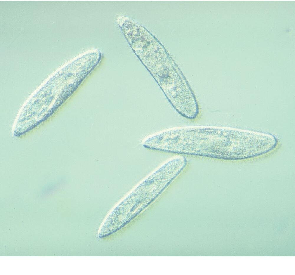
A very large barrel-shaped ciliate that feeds on Paramecium.

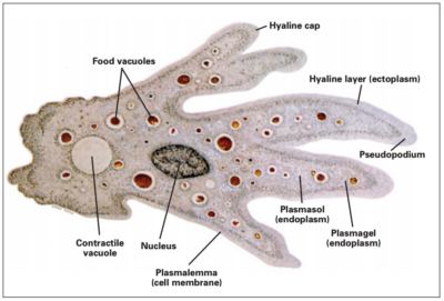
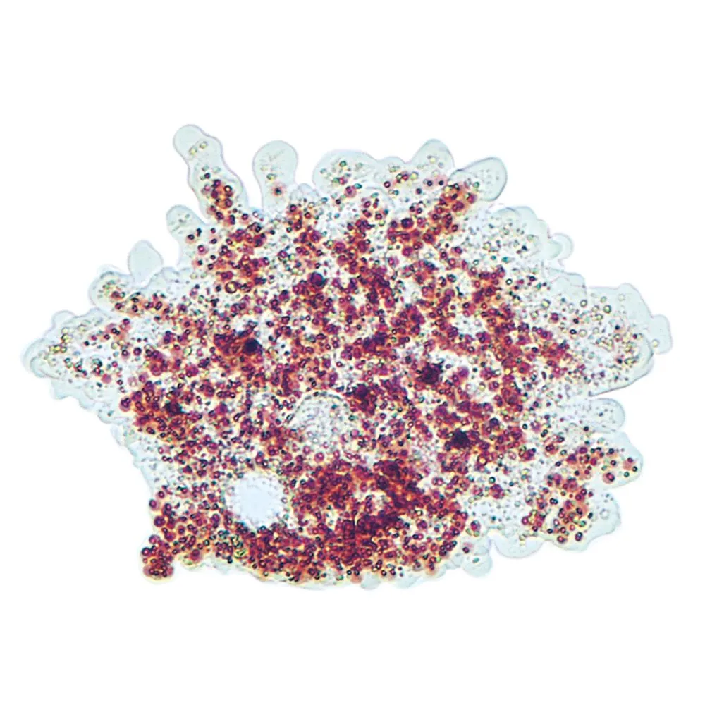
Also known as Pelomyxa, Chaos looks like Amoeba but us much larger and has many nuclei.

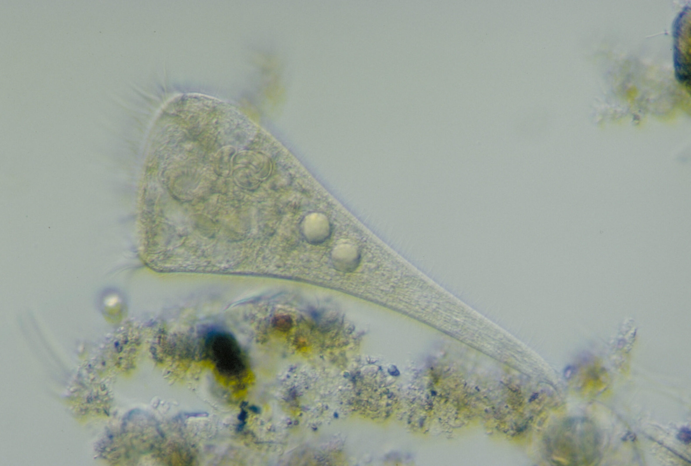
A bizarre ciliate with a long, tapering proboscis equipped with toxicysts that enable it to capture prey.

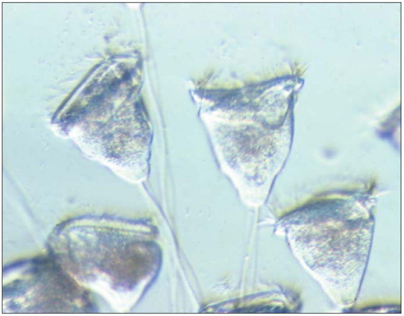
One of the most complex of the ciliates. Many of its cilia are bundled into leg-like appendages. An interesting lab activity is to pair Euplotes with a rotifer and ask, “Which is the animal, and how do you know?”

A marine, bioluminescent dinoflagellate. Under proper conditions, Pyrocystis produces a striking blue light. Highly recommended for studies of bioluminescence.
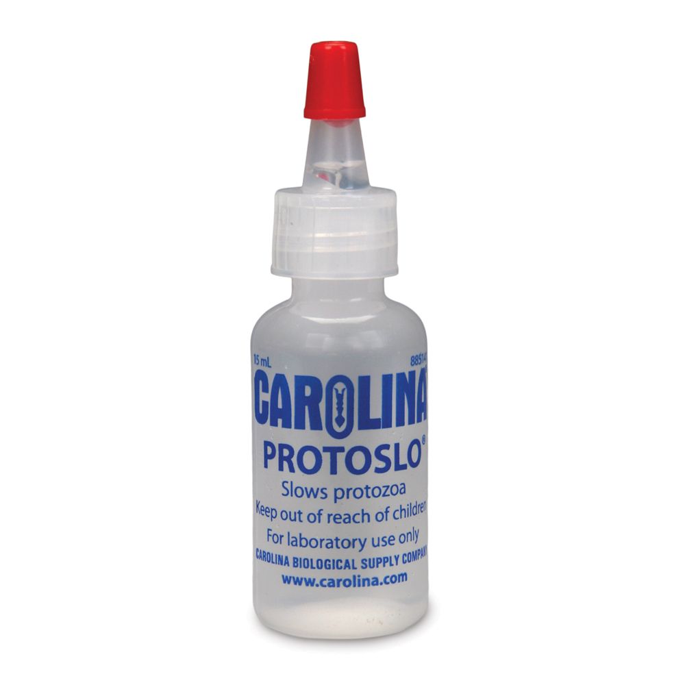
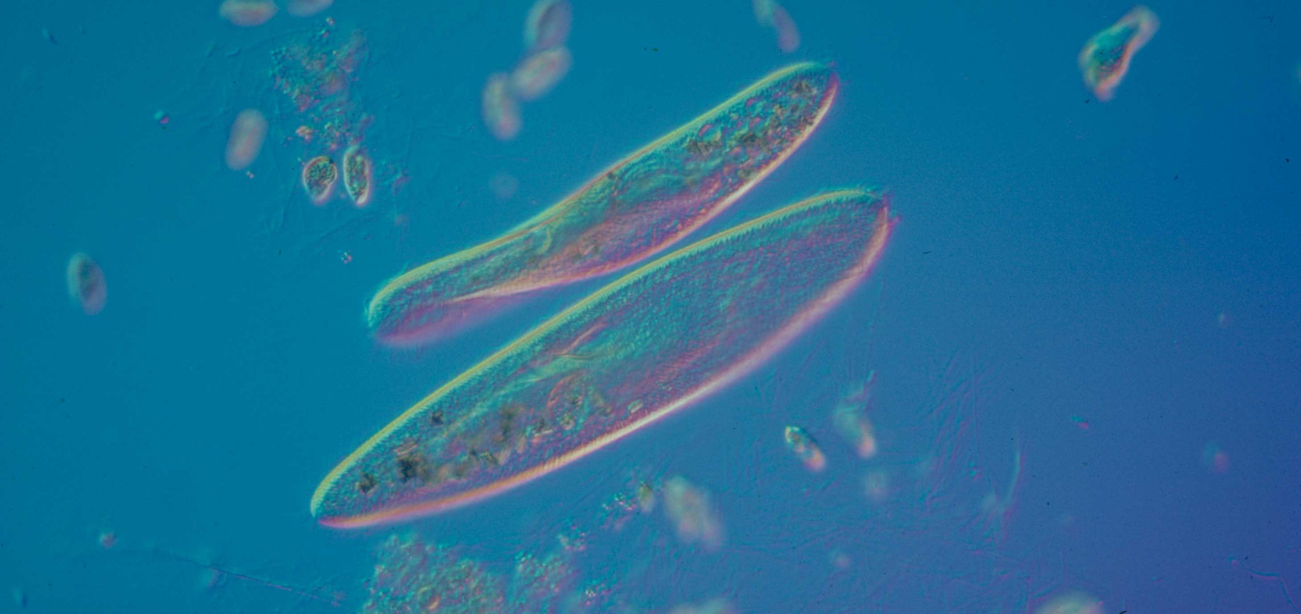
A microscope magnifies not only the size but also the apparent speed of living, motile subjects. Ciliates may zoom through your field of view. Protoslo slows them for better viewing at higher magnification.
A vital stain is one that does not kill the organism yet makes cytoplasmic structures more visible. Available for Amoeba, Chaos (Pelomyxa), and Paramecium cultures.

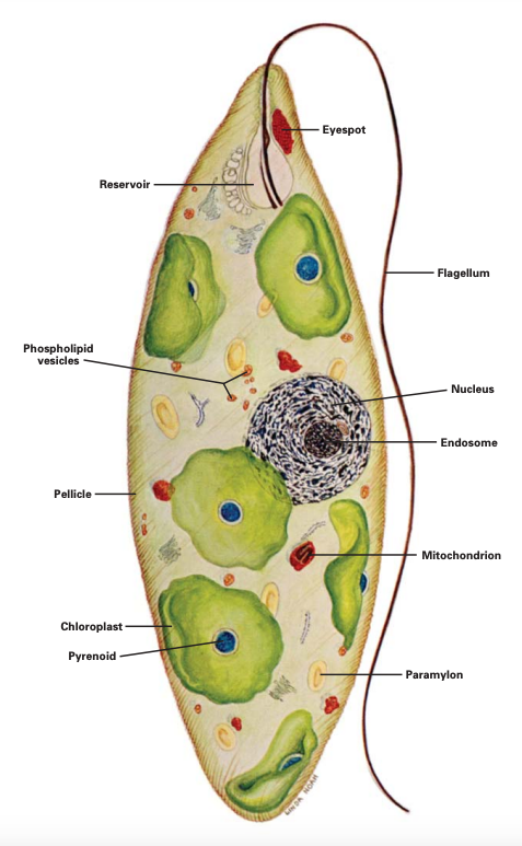
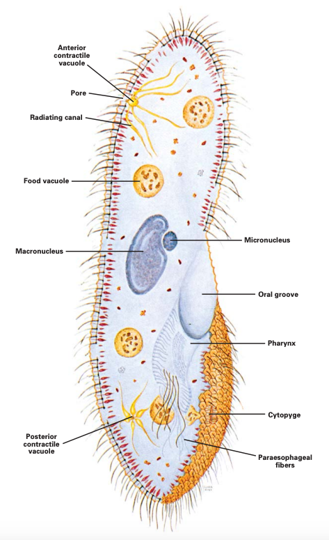
Pairs a live culture with a prepared, stained microscope slide of the protozoan to highlight internal structure. Available for Amoeba, Euglena, and Mixed Protozoa.


