CTE Health Science Education provides a comprehensive program to meet current and projected needs for the healthcare industry in fields including first aid, nursing, biomedical technology, and allied health care fields. Foundational curriculum concepts incorporate technological advances to motivate students, preparing them for a career as a future health professional. The domains of healthcare and related skills such as employability skills, prevention and wellness, diagnostics, therapeutics, and rehabilitation are emphasized in each career pathway.
This buying guide was developed to assist teachers with hands-on labs, materials, and content for the foundational course of Health Science I, as it is a gateway course for career pathways. This course focuses on human anatomy, physiology, and the human body diseases and disorders, and recognizing and responding to first aid emergencies. Students learn about healthcare careers within the context of human body systems.
Equipment and PPE
Suggested Equipment and PPE
The following equipment and PPE linked below may be needed for particular items listed in the buying guide.
706336A Nitrile Disposable Gloves
646852 Disposable Protective Face Mask, Pack of 50
706244 Lab Aprons
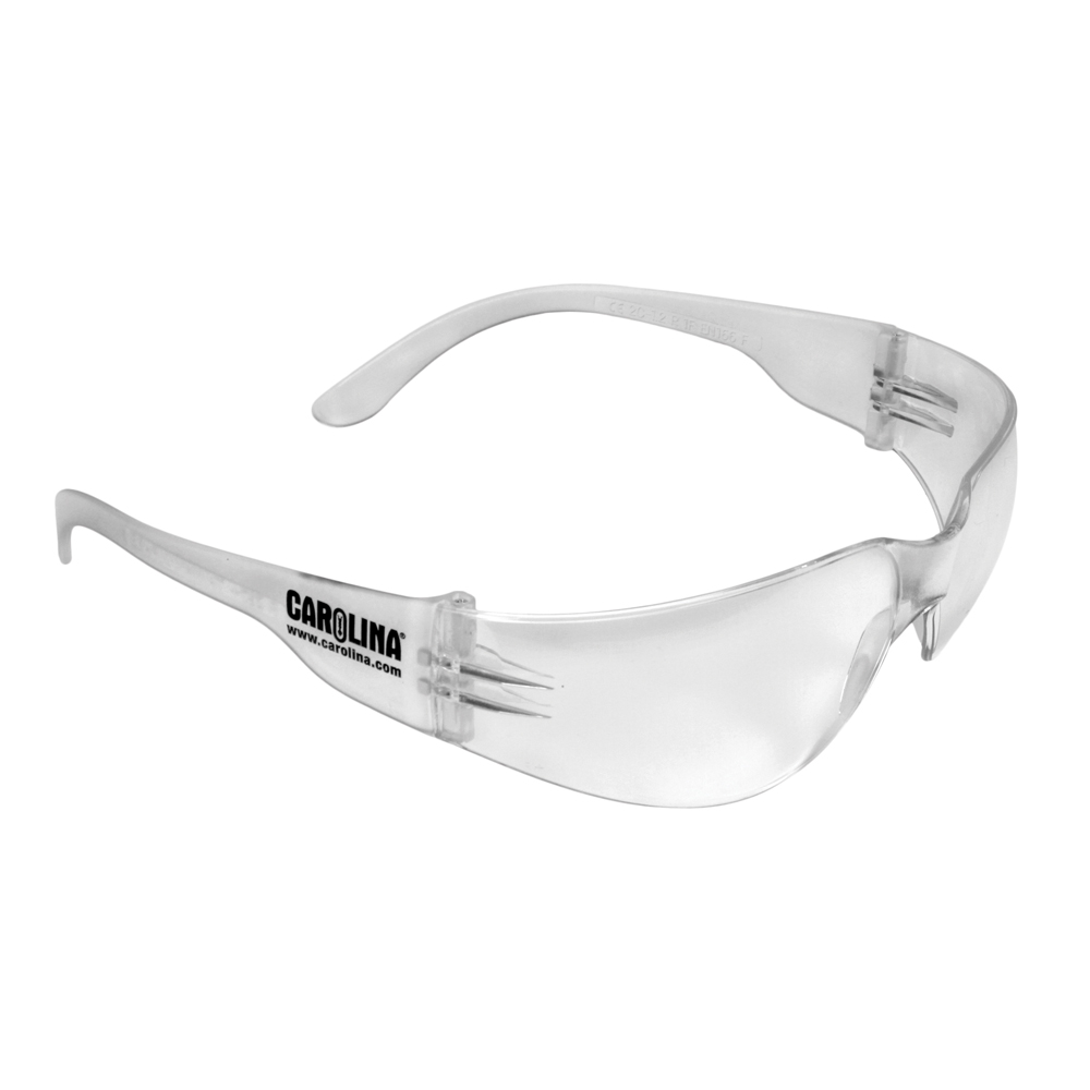
646700 Carolina® Safety Spectacles
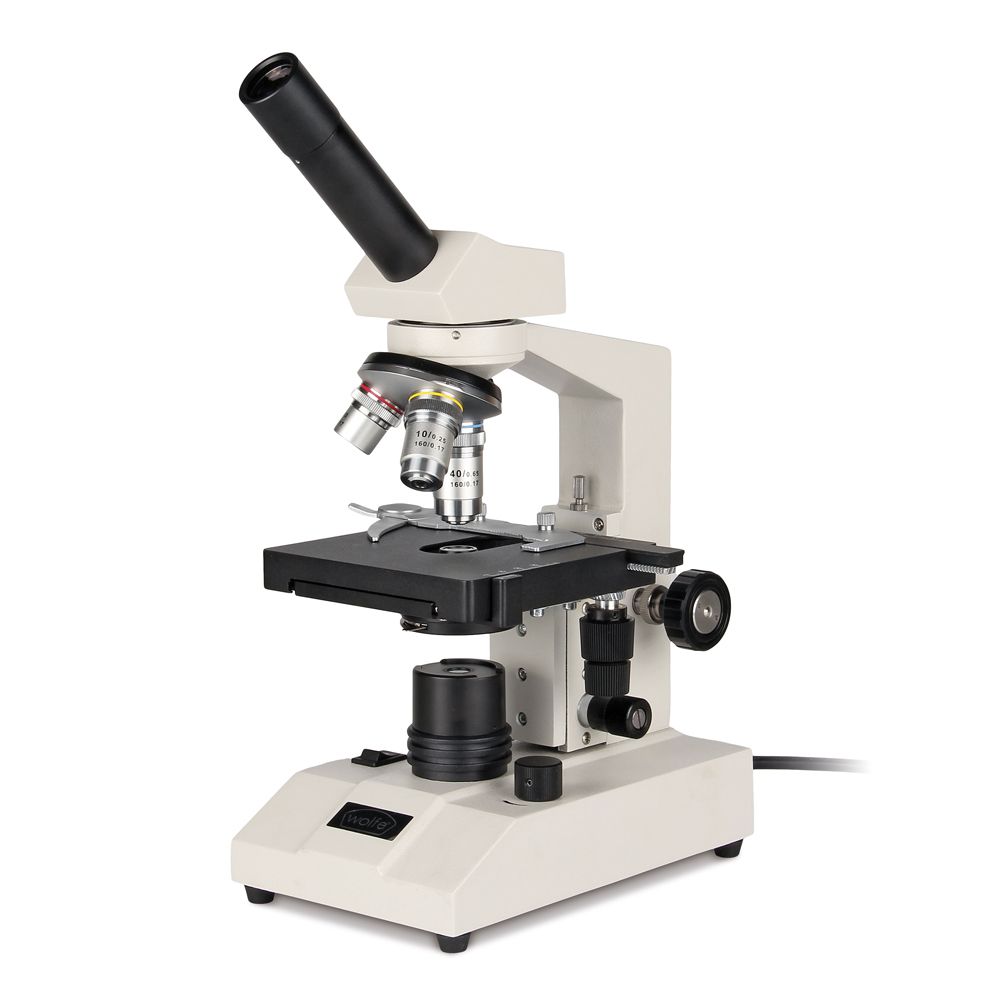
590945 Wolfe® HS Series Microscope with Mechanical Stage
591316 Wolfe® DigiVu™ DVM 6.0 Digital Microscope
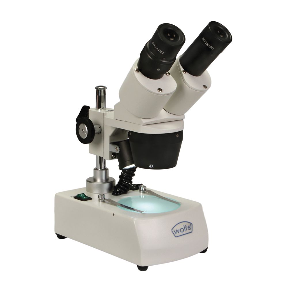
591810 Wolfe® Student Stereomicroscopes
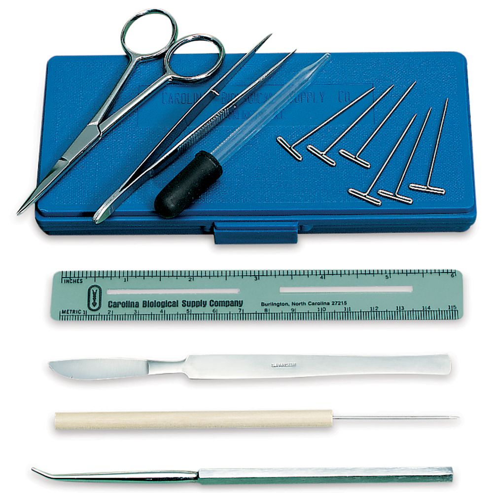
621176 General Biology Dissecting Set
Structural Organization
Anatomical Structural Organization
Before studying the various structures and functions of the body, learners need to analyze and understand the basic levels of structural organization that build upon one another. The five levels include: chemical, cellular, tissue, organ, and organism.
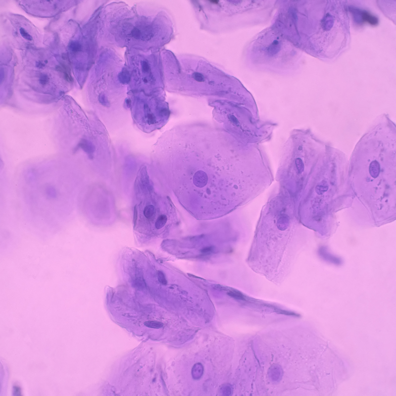
Human epithelial cheek cells
201100 Carolina BioKits®: Molecules of Life
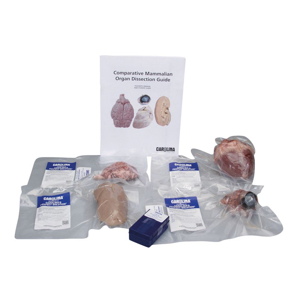
227975 Comparative Mammalian Organ Dissection Kit
292104 Discovering Animal Cells Self-Study Unit
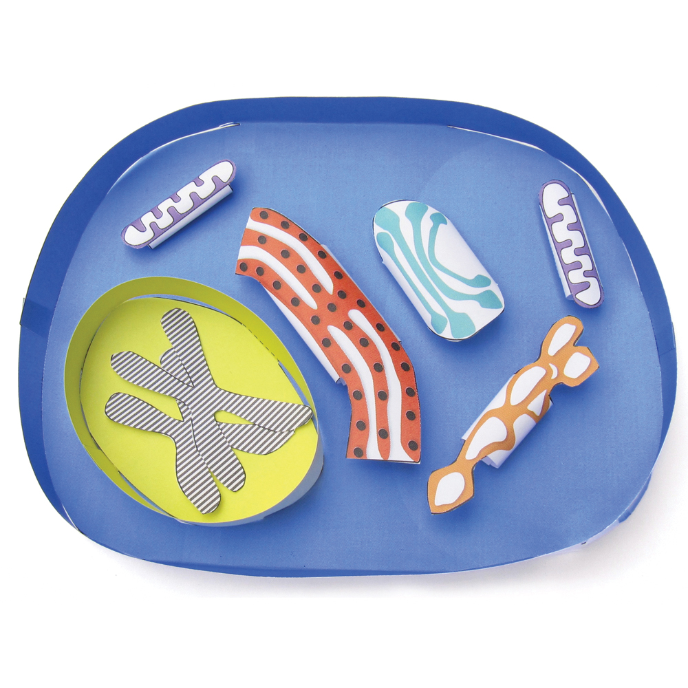
569650 3-D Paper Model Kit: Animal Cell
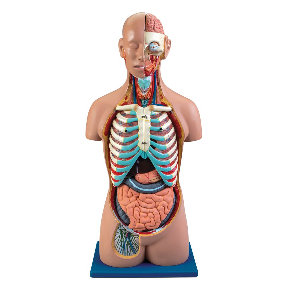
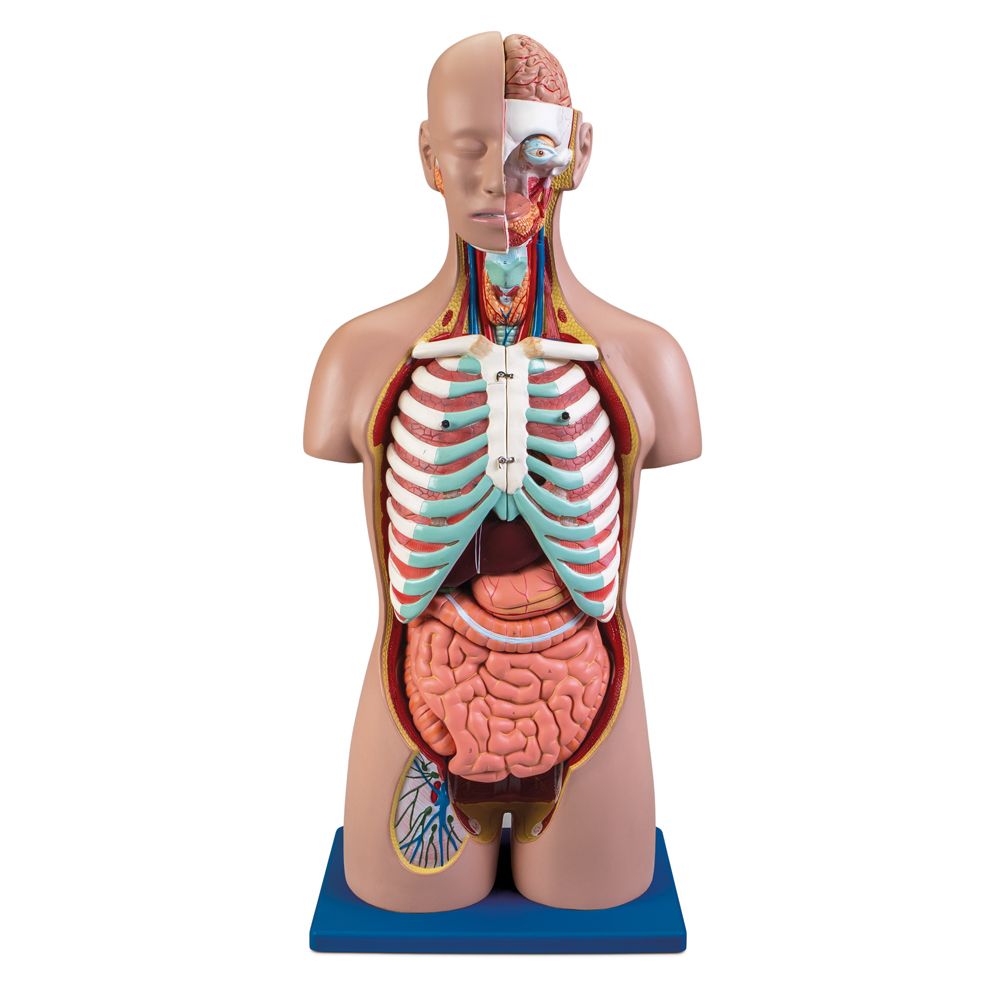
569520 Carolina® Human Sexless Torso Model with Open Back
567500 Life/form® Basic Buddy® and Baby Buddy® Classroom Pack
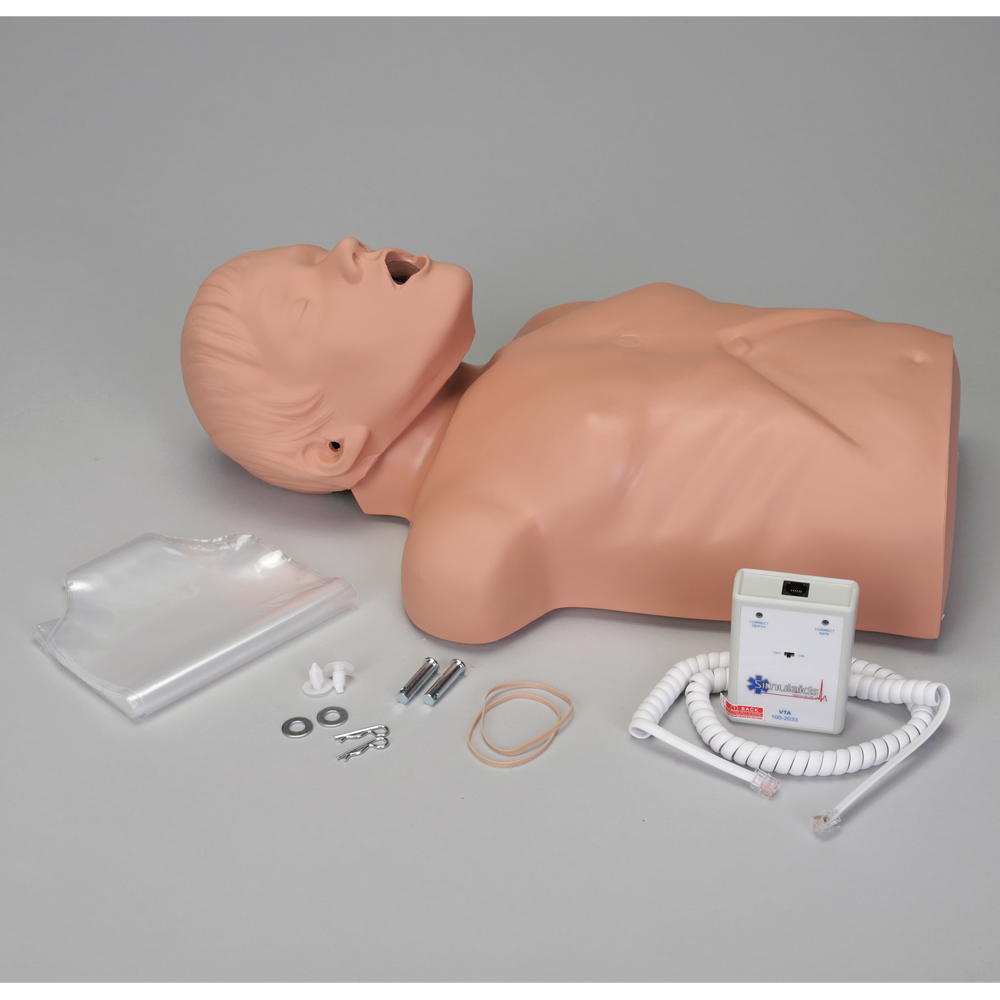
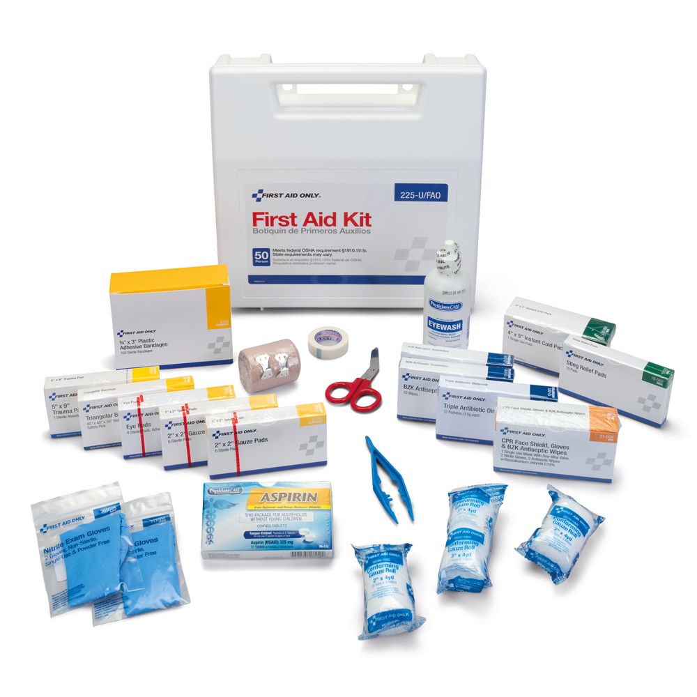
646514 Deluxe OSHA-Compliant First Aid Kit
691032 Blood Pressure Classroom Pack
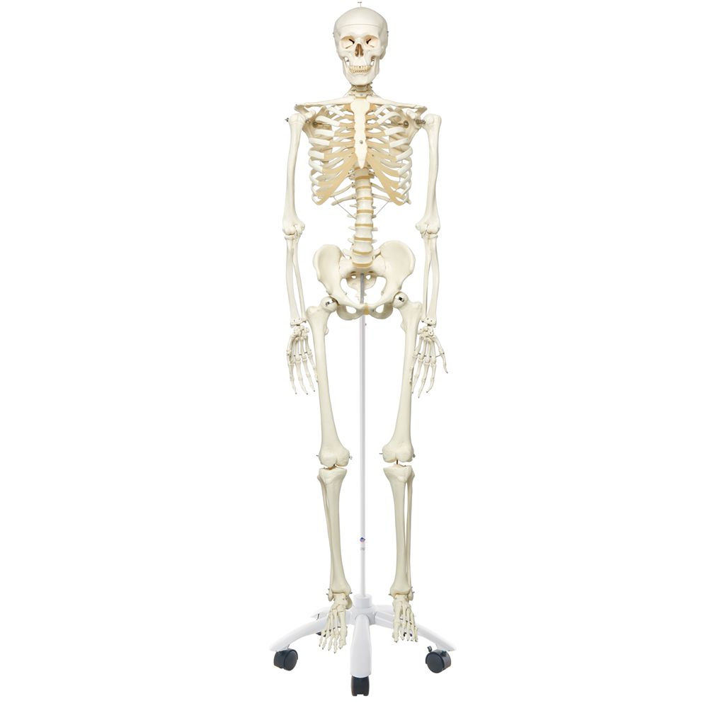
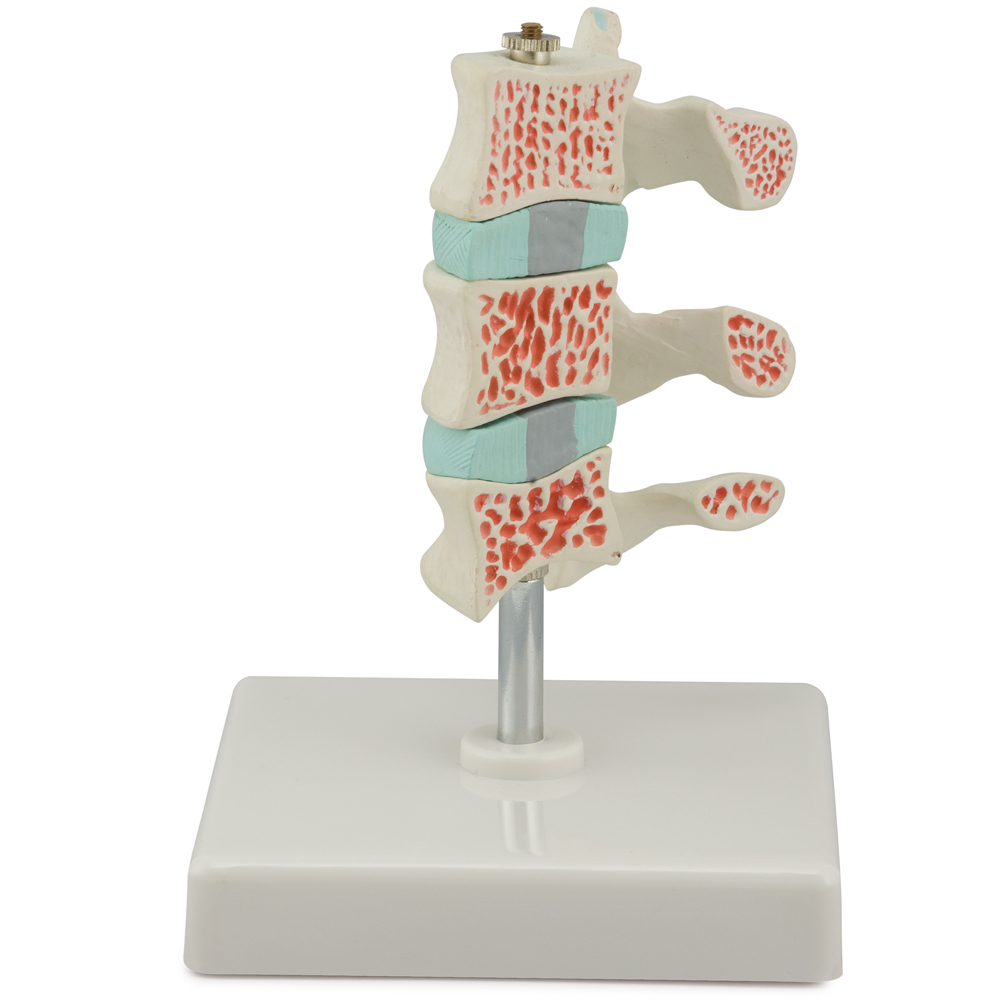
Skeletal System
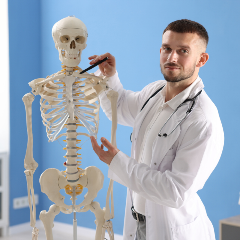
Skeletal System
The skeletal system comprises the skeleton, bone tissue, and cartilage. The skeleton is responsible for assisting with movement and protecting organs and soft tissue. Learners analyze and understand the structures, functions, and disorders of this system.
Doctor showing a human skeletal system model in a classroom.
Self-Adhesive Human Anatomy BodyPartCharts™
3B® Human Skeleton, Rod-Supported
Altay® Human Advanced Osteoporosis Model
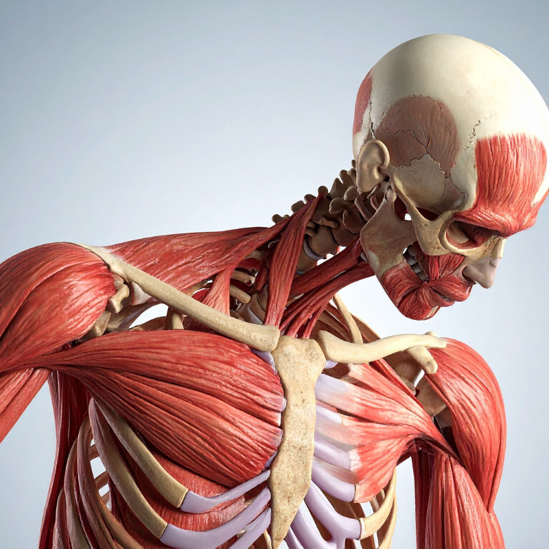
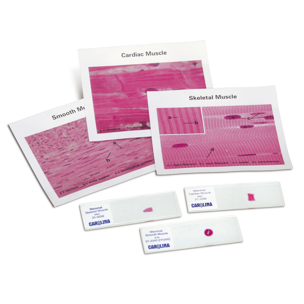
Muscular System
Muscular System
The muscular system is comprised of 3 types of muscle: smooth, skeletal, and cardiac. Not only does this system enable movement of the body, but it also regulates breathing, circulation, and digestion. Learners analyze and understand the structures, functions, and disorders of the muscular system.
Human muscular system
312058 Muscle Types Slide Set
566643A 3B Human Muscular Male and Female Model
203526 ATP Muscle Kit
229907 Carolina® Muscles of the Cat Dissection Mat
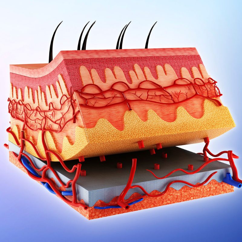
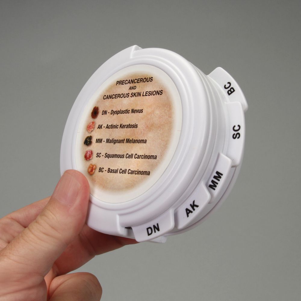
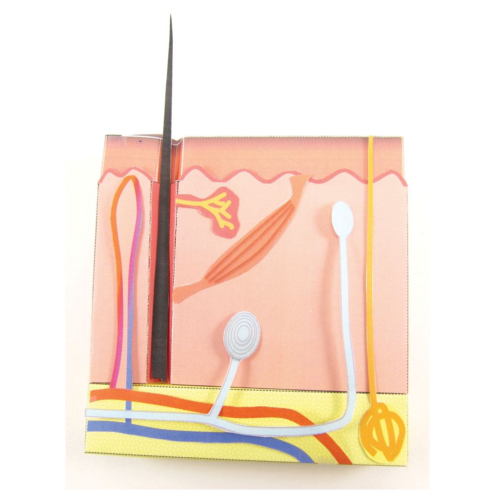
Integumentary
Integumentary System
The integumentary system is the largest organ system of the body and interfaces with the external environment. It consists of the skin, hair, and nails. Learners analyze and understand the structures, functions, and disorders of this system.
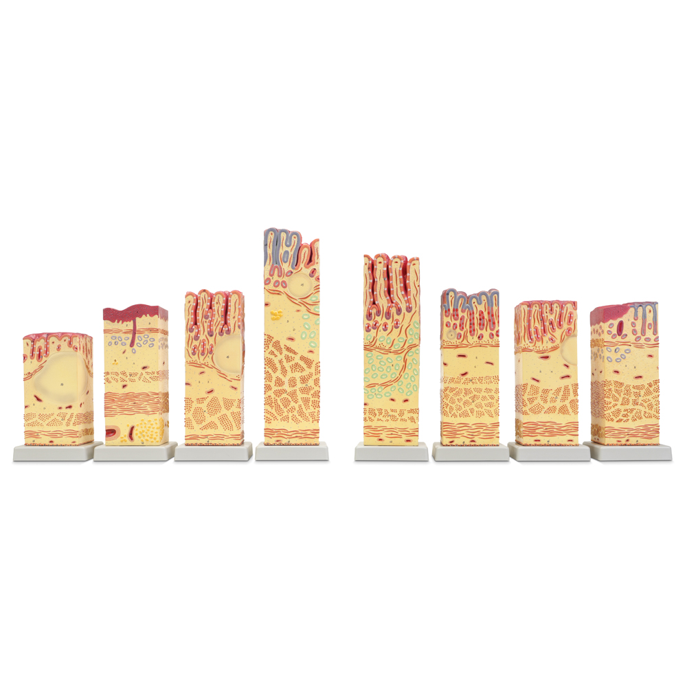
Cross section of human skin
Carolina® Enlarged Skin Model
GPI Anatomicals® Human Skin Cancer Disk Set Model
Origami Organelles™ 3-D Human Model Kits
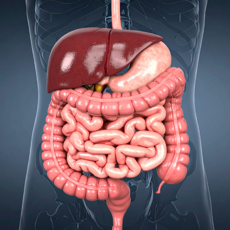
Digestive
Digestive System
The digestive system has multiple organs such as the stomach, liver, and small intestine. It is responsible for breaking down food into smaller molecules so that nutrients can be easily absorbed. Learners analyze and understand the structures, functions, and disorders of this system.
Human digestive system
202340 Carolina BioKits®: Digestion
Somso® Human Digestive Tract Model (item #566906)
566745 Altay® Human Comparative Digestive Tissue Model
576660 Giant Carolina® Human Anatomy Series Charts
GPI Anatomicals® Human Colon Pathology Model (item #569034)
Urinary
Urinary System
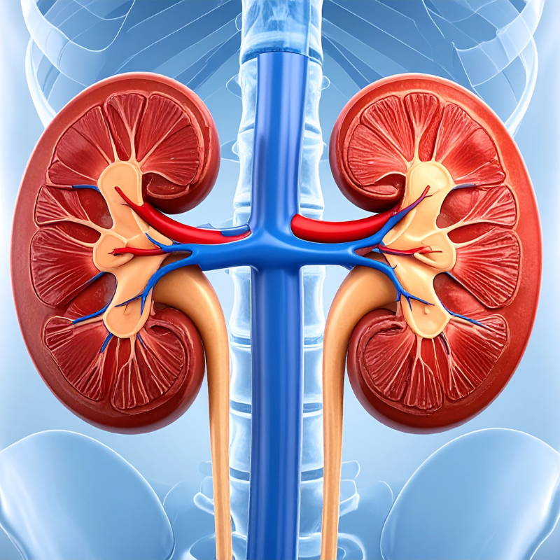
The function of the urinary system is to maintain proper fluid balance inside the body. The kidneys intertwine with arteries and veins to filter various waste products, such as salts and proteins, from the bloodstream. Learners will analyze and understand the structures, functions, and disorders of this system.
Illustration of human kidneys and urinary tract.
569170 GPI Anatomicals® Human Kidney Pathology Model
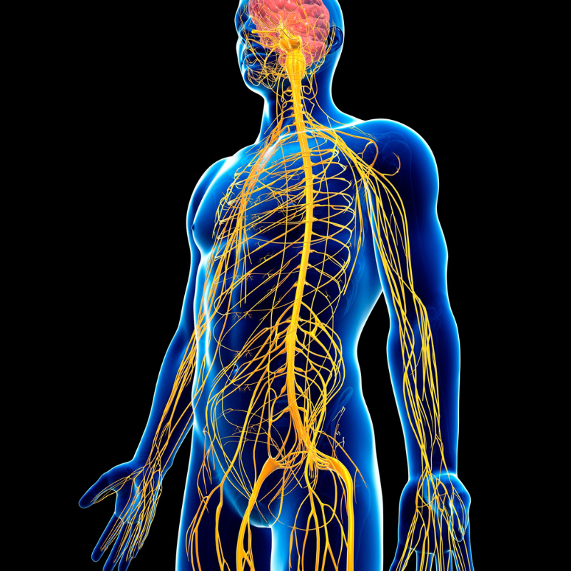
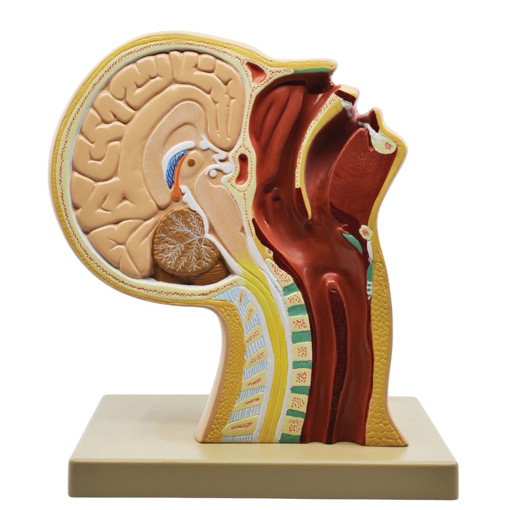
The nervous system is made up of the central nervous system and peripheral nervous system. Working together, they collect and interpret data from the body’s internal and external environments. Learners will analyze and understand the structures, functions, and disorders of this system.
Illustration of human nervous system.
569422 Carolina® Human Brain with Arteries Model
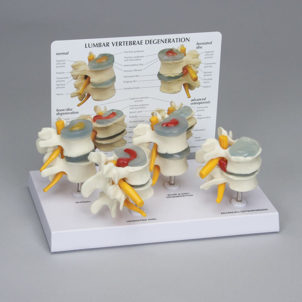
569200 GPI Anatomicals® Human Vertebrae Pathology Model Set
229910 Carolina® Brain Dissection Mat
566799 Altay Human Vertebrae Spinal Cord Dissection Model
566599 GPI Anatomicals® Human Brain Model in Skull with Pathologies
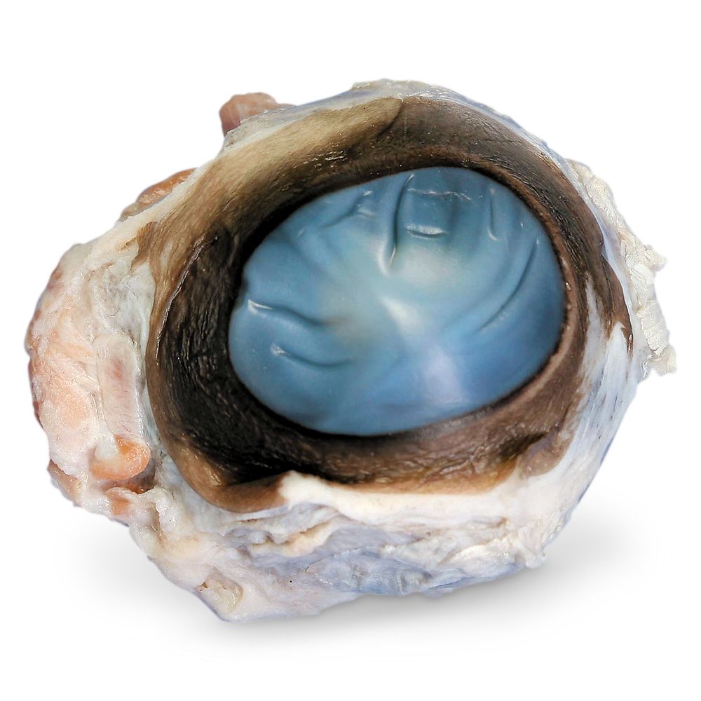
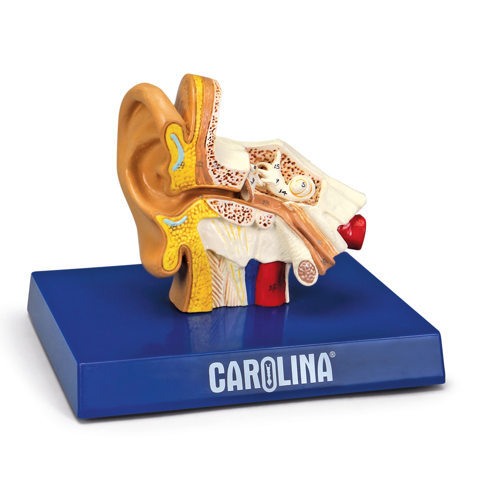
Sensory
Sensory System
Senses gather information from the external environment for processing by the brain, helping organisms survive. The sensory system is made up of the 6 senses that are specialized to detect a particular stimulus. Learners will analyze and understand the structures, functions, and disorders of this system.
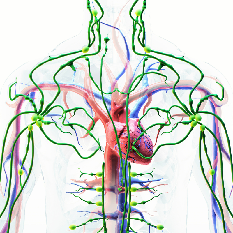
The lymphatic system plays a vital role in the body’s immunity to disease and pathogens. It’s responsible for allergic reactions as well as fighting diseases that range from the common cold to cancer. Learners will analyze and understand the structures, functions, and disorders of this system.
Human lymphatic system
Somso® Human Lymphatic System Model
Origami Organelles™ 3-D Paper Model Kit: Immune System
Endocrine
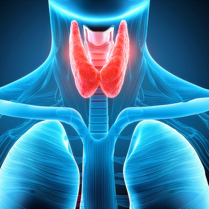
Endocrine System
The human body’s internal physiological control is provided by the endocrine system, which produces a variety of hormones responsible for regulating processes such as growth, heart rate, blood pressure, the sleep-wake cycle, metabolism, and reproduction. Learners will analyze and understand the structures, functions, and disorders of this system.
566746 Altay® Pathology of the Human Thyroid Model
566743 Altay® Pathology of the Human Pancreas, Duodenum, and Gallbladder Model
566908 Somso® Human Spleen, Duodenum, and Pancreas Model
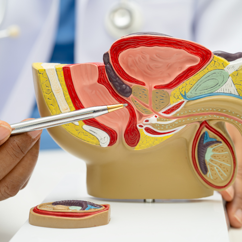
Reproductive
Male & Female Reproductive Systems
The male and female reproductive systems are controlled by hormones produced by the pituitary gland and the reproductive organs themselves. Organs in the reproductive system make, mature, and store gametes, which provide genetic variation through sexual reproduction. Learners will analyze and understand the structures, functions, and disorders of this system.
Anatomical model of male reproductive system.
Carolina® Female Half Pelvis Model (item #569470)
569471 Carolina® Male Half Pelvis Model
228360 Reproductive Rat Anatomy Kit
569100 GPI Anatomicals® Human Uterus and Ovary Pathology Model
569120 GPI Anatomicals® Male Pelvis with Testicular Pathology Model
229970 Carolina® Rat Dissection Mat
Carolina has the kits and supplies needed for your CTE students to master the essential knowledge and workplace skills required for careers in the healthcare industry.
Don’t see a specific item or content area covered that you need for your CTE Health Science I course? Contact us for special orders! We stand by our 100% customer satisfaction guarantee and will work with you to find what your students need.