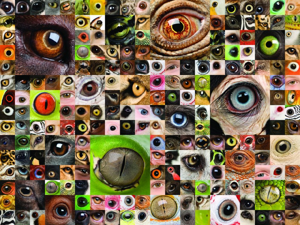
The light-sensing organs and eyes of living organisms have long fascinated the general public and scientists alike. Scientists ask questions such as “How do the ‘eyes’ on planaria work?,” “What is the ‘eyespot’ on the Euglena for?,” and “What are the steps in the development of zebra fish vision as the animal develops from a fertilized egg to an adult fish?” They study light-responding organs to seek answers to those questions and to:
- Understand how the organs work
- Discern the evolutionary relationships between them
- Gain a greater understanding of human eye disease by studying simpler models
The following is a brief overview of the increasing complexity found in the light-sensing organs of organisms as they become physiologically more complex. It also briefly describes the structure of light-sensing organs and/or eyes in several diverse organisms that can be studied in the classroom.
Evolutionarily, photoreception is thought to have begun with the simple perception of light vs. dark. This perception allowed an organism to sense and respond to circadian rhythms in the environment, and to a certain degree orient itself in its surroundings. The next level of complexity displayed by light-sensing organs was the ability to detect the direction from which light came. This ability allowed an organism to orient itself with more specificity within its environment. Organisms such as planaria and Euglena gracilis have these types of light-sensing organs. Finally, the development of true spatial vision occurred. The development of this kind of vision allowed an animal to see images and judge distance. Zebra fish have this type of visual ability. True spatial vision enables more complex interactions with the environment, such as those involved in hunting prey.
Two different categories of eye developed that conferred spatial vison. One type is the compound eye, such as those seen in insects. Compound eyes have multiple lenses or receptors that send light to a small number of photoreceptors that then process the images. The second type of eye is the camera eye found in most vertebrates, such as humans, and in cephalopods, such as the squid. A camera eye contains a single lens that focuses incoming images onto a sheet of photoreceptors at the back of the eye (the retina). The image is then sent to, and processed by, the brain.
The light-detecting systems of 3 organisms are briefly described below. These descriptions should provide you with an idea of the range in complexity displayed by the visual systems found in living organisms. They should also allow you to see the connections between the different visual systems.
Euglena gracilis


Euglena are single-celled protists that live in shallow ponds. They obtain food either by absorbing nutrients or through photosynthesis using their chlorophyll. Euglena are able to perceive light and the direction it comes from through the use of 2 organelles, an eyespot and a photoreceptor. The 2 organelles function in conjunction with the organism’s pattern of movement. The Euglena uses information about the direction light comes from so it can orient itself to maximum advantage in the environment. For example, when it needs to use photosynthesis to generate food it can position itself to optimally obtain the needed energy from sunlight.
Euglena’s red eyespot is composed of carotenoid pigments. Its photoreceptor is composed of multiple layers of protein-imbedded membranes. The protein found in the membranes is, within each layer, arranged in a hexagonal lattice structure and belongs to the rhodopsin family. This arrangement of the proteins forms a type of crystalline structure that is thought to optimize the amount of light detected by the photoreceptor.
The rhodopsin protein in the photoreceptor has 2 different forms. The dominant form of the protein absorbs in one range of wavelengths. When the protein absorbs light in that range, it changes to the other conformation. This second conformation is the signaling state of the protein. When it is in this state, the protein alters the structure of the flagella the organism uses to move and thus changes the organism’s movement. A different range of wavelengths of light cause the photoreceptor protein to reset back to its non-signaling state.
The organism detects the direction from which light comes by continuously moving in a helical pattern and thus sampling the environment. At certain points in this movement, the Euglena’s eyespot blocks the wavelengths of light that convert the protein back to the more dominant, non-signaling form and prevents them from hitting the photoreceptor. The resulting shift in the ratio of the 2 forms of the photoreceptor alters the movement of the organism.
Planaria

Planaria are nonparasitic aquatic flatworms (Platyhelminthes) that have a simple nervous system with a brain and are known for their ability to regenerate. These animals avoid light, especially ultraviolet (UV) light, presumably to hide from predators and to escape harm from UV light exposure. The animal detects light and the direction it comes from by using the 2 simple eyes on its head. Planaria’s eyes contain 2 different types of cells: pigment cells that form a half-moon-shaped cup that absorbs the incoming light, and photoreceptors that transmit the information collected by the pigment cells to the animal’s simple brain.
The pigments in the pigment cells are rhodopsins and change their conformation in response to light in a way similar to that of the rhodopsins in the Euglena. The cell bodies of the photoreceptors lie outside of the cup, with their dendrites, branches of the cell for receiving signals, extending into the cup. The axons of the photoreceptors enter the brain and thus transmit the signals they receive for processing by the brain.
Planaria can perceive the direction of incoming light partly because of the shape of the eye cup. Light hits different parts of the eye cup depending upon the direction from which it comes. In addition, the 2 eyes are on different sides of the animal’s body and thus receive different visual information. The animal’s simple brain processes the different information coming from the 2 sides to help distinguish where the light is coming from.
Scientist have used planaria’s avoidance of light to study the animal’s ability to regenerate. If its head is removed, the animal no longer avoids light, presumably because it can no longer sense it. As the animal regenerates its head, eyes, and brain, it regains its sensitivity to light. By correlating when the animal regains its sensitivity to light with the reappearance of various structures and proteins, researchers can learn about the steps involved in the regeneration of this sensory ability.
Zebra fish (Danio rerio)
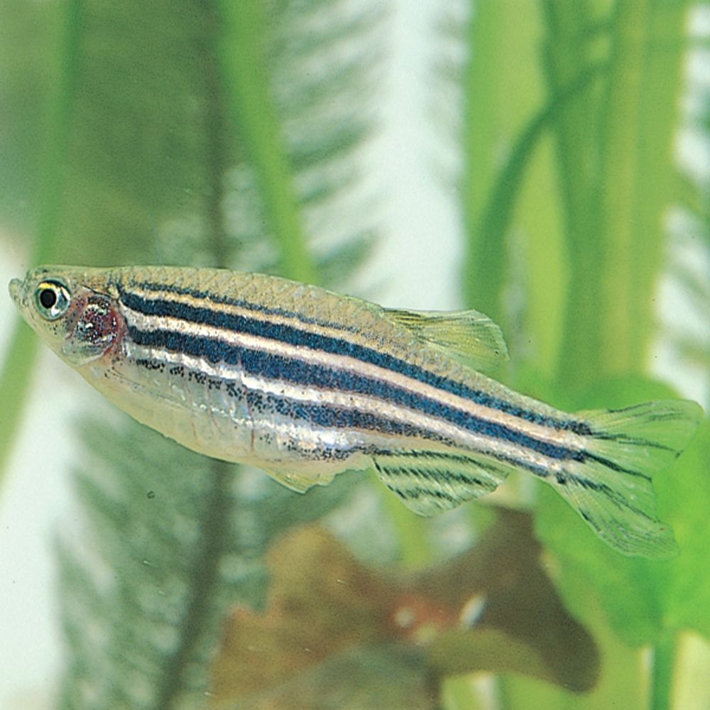
The zebra fish eye is, like human and other vertebrate eyes, a camera eye. It contains a single lens that focuses images onto a retina. The zebra fish eye has a cornea that protects it from the environment. The cornea is the transparent tissue that covers the eye and is contiguous with the sclera, the tissue making up the tough outer shell of the entire eye. The sclera helps maintain the eye’s structure and protects it.
The lens sits behind the cornea and focuses images on the retina. The retina of the zebra fish, like the human retina, is composed of multiple layers of cells, both neurons and photoreceptors. Like human eyes, zebra fish eyes have 2 major categories of photoreceptors: rods and cones. Zebra fish cones operate in bright light, and the rods in low light.
Like human eyes, zebra fish eyes have cones that respond to red, green, and blue light. However, in the zebra fish, the red and green receptors are merged. Zebra fish also have photoreceptors that can detect UV light. The ability of the different photoreceptors to detect different wavelengths of light is linked to the type of pigment each contains. The complex array of neurons in the retina and in the animal’s brain process the information from all these diverse receptors into meaningful information for the animal.
The importance of these neurons to the animal’s vision may explain why experiments indicate that young zebra fish that seem to have all the necessary anatomy for full-color vision do not appear to be able to see in color. The hypothesis is that further neural development needs to occur before the fish can obtain full-color vision. This explanation is consistent with earlier observations that zebra fish’s vision continues to develop even after they hatch.
Zebra fish are what is called a model organism in biological research. They are similar enough to more complex organisms (like humans and other vertebrates) that studying them provides scientists with insight into how the more complex organism works. They can be used to perform experiments geared to understanding human vision. For example, in humans, hereditary blindness is often caused by mutations that lead to degeneration of the photoreceptors. Zebra fish have similar types of mutations. By studying these mutations and their effects in zebra fish, scientists can learn how the mutations affect human vision.
Other interesting observations about zebra fish that investigators have noted are listed below. Scientists have taken advantage of some of these behaviors when designing experiments.
- Zebra fish will reflexively follow a moving striped pattern. This behavior appears around the time they hatch (73 hours after the eggs were fertilized) and continues to be refined until about 96 hours after fertilization.
- Zebra fish raised in continuous light and those raised in continuous dark exhibit different visual behavior from those raised around normal light patterns. Their behavior returns to near normal once their lighting conditions are returned to normal.
- Data suggests that zebra fish completely turn off their visual functions at night.
The information given here is just a fraction of the information known regarding these organisms and their light-sensing abilities. It is hoped that it has given you an idea of the variation and complexity by which organisms sense and respond to light in their environment, and that some of the information will inspire you to design your own experiments in the classroom.
Barsanti, L., Evangelista, V., Passarelli, V., Frassanito, A.M., Gualtieri, P. 2012. Fundamental Questions and Concepts about Photoreception and the Case of Euglenia gracilis. Integrative Biology 4, 22–36.
Bilotta, J., Saszik, S. 2001. The Zebrafish as a Model Visual System. International Journal of Developmental Neuroscience 19, 621–629.
Gestri, G., Link B.A., Neuhauss S.C.F. 2012. The Visual System of Zebrafish and Its Use to Model Human Ocular Diseases. Developmental Neurobiology 72(3), 302–327.
Inoue, T., Kumamoto, H., Okamoto, K., Umesono, Y., Sakai, M., Alvarado, A.S., Agata, K. 2004. Morphological and Functional Recovery of the Planarian Photosensing System During Head Regeneration. Zoological Science 21, 275–283.
Lamb, T.D. 2011. Evolution of the Eye. Scientific American (July): 64–69.
Nilsson, D. 2009. The Evolution of Eyes and Visually Guided Behavior. Philosophical Transactions of the Royal Society B. 364, 2833–2847.
Paskin, T.R., Jellies, J., Bacher, J., Beane, W.S. 2014. Planarian Phototactic Assay Reveals Differential Behavioral Responses Based on Wavelength. PLOS ONE (December): DOI:10.1371/journal.pone.0114708.
Yong, E. 2016. Seeing the Light. National Geographic (February): 38–57.